


Transmission Electron Microscopy: New Advances and Applications for Earth and Planetary Sciences
-
摘要: 近年来,各种微束分析技术的快速发展及其在地球和行星科学领域的广泛应用,极大地推动了纳米地球科学和行星科学的学科发展和科学研究.透射电子显微镜(简称透射电镜)因具有空间分辨率高和综合分析能力强等优点,在地球与行星物质的微纳尺度到原子水平的形貌、晶体结构、矿物相鉴定、化学成分、原子成像和微磁结构等研究中发挥着巨大作用.在简要回顾透射电镜的发展历程、物理结构和工作原理的基础上,结合本实验室过去几年的工作内容,重点介绍了透射电镜的基本功能、样品制备方法及其在地球和行星科学研究中的应用范例.通过与其他微束分析技术的简单对比,还初步分析了透射电镜在地球与行星科学研究领域的应用现状和未来趋势.Abstract: In recent years, researches on nanogeoscience and planetary sciences have achieved significant progresses mostly due to the applications of various microscopic and micro-spectroscopic approaches. Among these techniques, transmission electron microscopy (TEM), known as high spatial resolution and strong comprehensive analysis capability, plays an important role in simultaneously imaging and characterizing morphological, structural, chemical, and micromagnetic properties down to atomic scales. In this review it briefly introduces the development history, mechanical structure and working principles of transmission electron microscope, and the sample preparation involved in the TEM technique as well. Based on some recent progresses achieved in the Electron Microscope Lab at the IGGCAS, this review also shows and focus on the fundamental functions and applications of transmission electron microscope in some typical researches in earth and planetary sciences. Finally, it tentatively discusses the current situation and future trend of transmission electron microscopy in earth and planetary sciences.
-

图 1 20世纪30年代初鲁斯卡(右)和克诺尔(左)研制的电子显微镜(Williams and Carter, 2009)(a);最初的电子显微镜光路图(b);现代球差校正透射电镜(JEM-ARM 300F,https://www.jeol.co.jp/en/products/detail/JEM-ARM300F.html)(c);聚光镜像差校正透射电镜结构示意图(d)
Fig. 1. The earliest electron microscope built by Ruska (right) and Knoll (left), in Berlin in the early 1930s(a), schematic illustration of the electron optical systems of the earliest electron microscope(b), picture of one representative modern transmission electron microscope (JEM-ARM 300F, https://www.jeol.co.jp/en/products/detail/JEM-ARM300F.html) (c), schematic illustration of aberration-corrected transmission electron microscope(d)
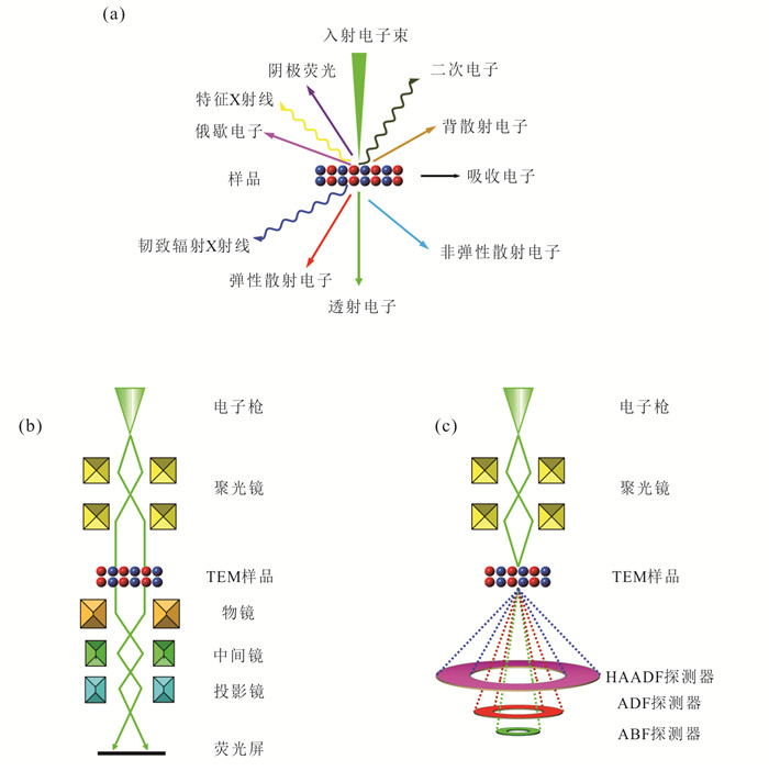
图 2 入射电子与样品相互作用后产生的各种信号(a);常规透射电子显微镜工作原理示意图(b);扫描透射电子显微镜工作原理示意图(c)
Fig. 2. Multiphoton absorption and emission by interaction between incident electrons and matters(a), schematic diagram of working principle of conventional transmission electron microscope(b), schematic illustration of working principle of a scanning transmission electron microscope(c)
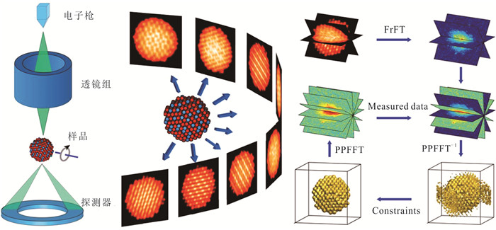
图 3 原子电子三维重构(AED)原理(Miao et al., 2016)
Fig. 3. The schematic layout of 3D atomic electron tomography (AET)
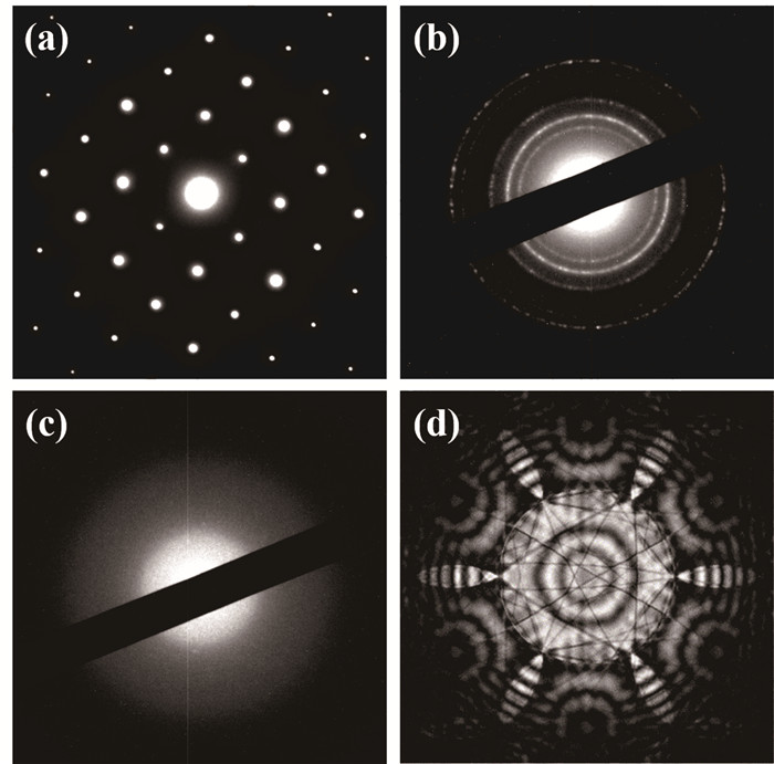
图 4 单晶衍射花样(橄榄石)(a);多晶衍射花样(Fe3O4)(b);非晶衍射花样(长石)(c);会聚束衍射花样(晶体材料)(d)
Fig. 4. SAED pattern for single crystal (olivine) (a), SAED pattern for polycrystal (Fe3O4) (b), SAED pattern for amorphous material (feldspar) (c), central disk of CBED pattern for the crystal model(d)
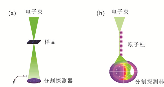
图 5 DPC STEM工作原理示意图(修改自Shibata, 2019)(a).分割探测器几何结构及其接收的电子束穿过原子柱时在衍射面产生的强度分布(修改自Sánchez-Santolino et al., 2018)(b)
Fig. 5. Schematic illustrations of DPC STEM(a), the segmented area detector geometry and the accepted intensity distribution in the diffraction plane when the electron probe passes close to a column(b)
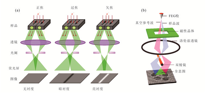
图 6 洛伦兹电子显微术的Fresnel成像原理(修改自Du et al., 2015)(a);离轴电子全息术原理(修改自Midgley and Dunin-Borkowski, 2009)(b)
Fig. 6. Schematic ray diagram in a Fresnel image of a ferromagnetic specimen containing two 180° domain walls(a), schematic diagram of off-axis electron holography (b)
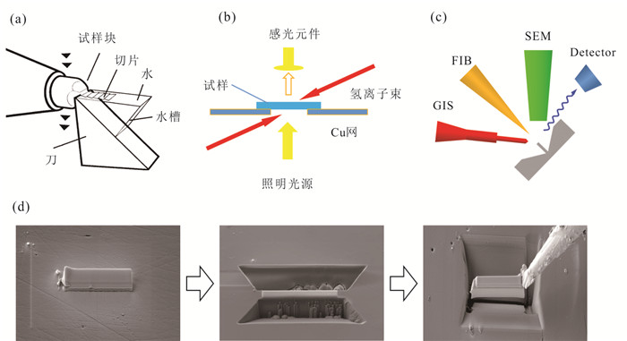
图 7 固体块状样品的透射电镜样品制备方法
a.超薄切片法工作原理图(Wei and Li, 1997);b.离子减薄工作原理图;c. FIB-SEM双束系统原理图;d. FIB-SEM制备TEM样品示意图
Fig. 7. Preparation methods of solid block sample for TEM
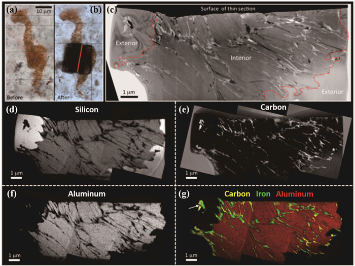
图 8 来自Apex燧石样品中的疑似细菌化石纳米结构和化学特征
图a和图b分别是提取用于TEM分析的超薄片之前和之后的光学显微照片.图c~f是伪化石薄片的明场TEM图像(c)和相应的TEM能量过滤像元素图,硅(d)、碳(e)和铝(f).图g为碳、铁和铝的元素叠加图,碳(黄色)和铁(绿色)交错于铝硅酸盐(红色)之间(Brasier et al., 2015)
Fig. 8. Nanoscale structure and chemistry of a pseudofossil from sample Apex chert

图 9 趋磁杆菌WYHR-1及其子弹头形磁小体晶体生长机制
a, b.一个WYHR-1细菌的明场像和高角环形暗场STEM图像;c.STEM-EDXS研究WYHR-1细胞的化学成分分布;d.磁小体链的STEM-HAADF三维层析重构图像;e, f.WYHR-1磁小体链的ACOM晶体取向分布图;e.对照指数图;f.水平方向(X)的晶体取向图;g.磁小体的晶体长度与宽度对比图及其磁小体晶体的生长规律;h, i.WYHR-1磁小体晶体的3D层析重构图(h)和晶体模型(i)(修改自Li et al., 2020d)
Fig. 9. Morphology, chemistry, crystal orientation and morphological model of WYHR-1 magnetosomal magnetite analyzed by atomic resolution STEM
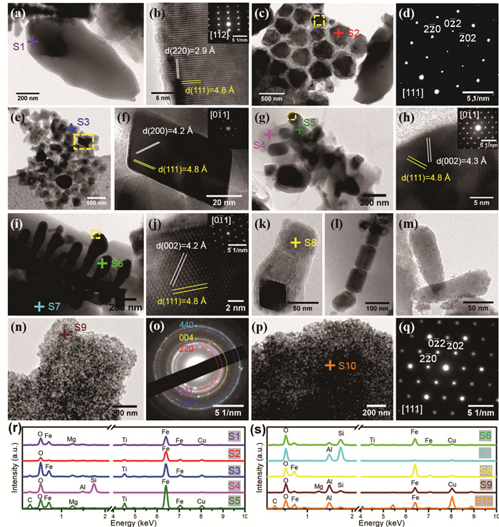
图 10 8种磁性矿物颗粒的TEM实验结果
a, b.类型1的单个磁铁矿颗粒TEM图像(a)和对应的高分辨像(b)以及电子衍射谱(图b插图);c, d.类型2的TEM图像(八面体的磁铁矿颗粒,平均粒径为367.8±44.9 nm)(c)和对应的电子衍射谱(d);e, f.类型3纳米级钛磁铁矿的TEM图像(e)和对应的高分辨像(f)以及电子衍射谱(图f插图);g, h.类型4的TEM图像(硅酸盐矿物包裹的纳米级钛磁铁矿)(g)和对应的高分辨像(h)以及电子衍射谱(图h插图);i, j.类型5的TEM图像(树枝状的磁铁矿颗粒)(i)和对应的高分辨像(j)以及电子衍射谱(图j插图);k, m.类型6的TEM图像(化石磁小体);n, o.类型7的超顺磁磁性矿物聚集体的TEM图像(n)和对应的电子衍射谱(o);p, q.类型8的TEM图像(取向一致的超顺磁磁性矿物的聚集体)(p)和对应的电子衍射谱(q);r, s.来自8种类型磁铁矿样品的EDXS成分分析,S1~S10表示图 10中十字叉位置(Li et al., 2020b)
Fig. 10. TEM results of eight types of magnetite mineral particles

图 11 卡林型金矿中纳米金的透射电镜表征
a.金粒子(较亮的点,箭头所指)的STEM-HAADF像;b~c.金晶粒的高分辨图像(HRTEM)(b)和对应的FFT图像(c);d~e.金粒子的EDXS元素面分布图,分别是Au -Lα (d)、Fe-Kα (e)和S-Kα (f)(修改自Palenik et al., 2004)
Fig. 11. Characterization of gold nanoparticles in a Carlin-type deposit by TEM
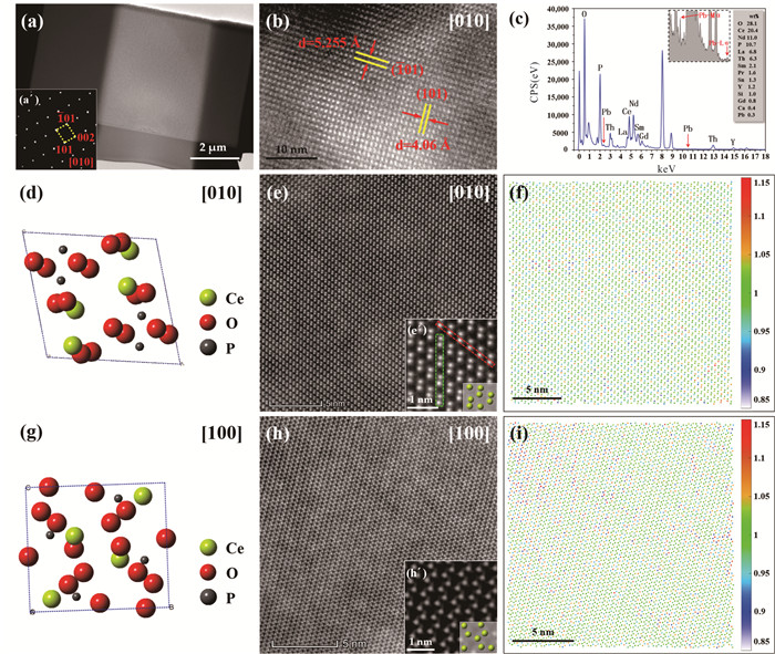
图 12 RW-1独居石中放射成因Pb的透射电镜分析
a.独居石薄片的TEM明场像和相应的电子衍射谱;b. [010]带轴的高分辨像;c.图a独居石晶粒的EDXS谱图,其中插图是放大的EDXS谱图,红色箭头分别指示Pb-Mα和Pb-Lα峰. d~i.沿着[010]方向(d~f)和[100]方向(g~i)的独居石原子模型(d,g)、HAADF像(e,h)和对应的归一化强度mapping(f,i);插图ex,hx分别是沿着[010]和[100]方向的放大的HAADF图像和相应的Ce原子模型,黄绿色球代表Ce原子;图f和i中白色-蓝色强度值(0.85)表示REEs分布,黄色-红色强度值(1.15)表示Pb和Th的分布(修改自Tang et al., 2020)
Fig. 12. TEM analysis of radiogenic Pb in RW-1 monazite
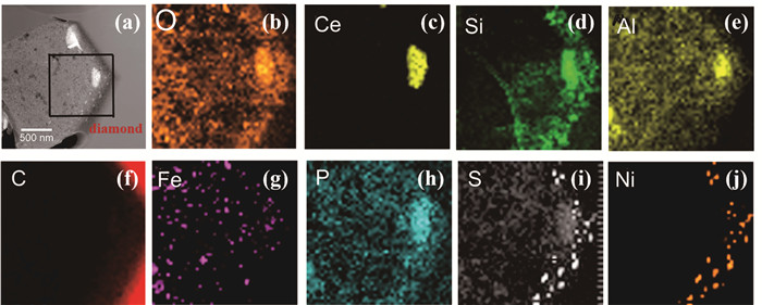
图 13 巴西Junia地区金刚石内多晶包裹体中集合体的STEM-HAADF图像(a)和EDXS元素分布(b~j)
Fig. 13. STEM-HAADF image (a) and elemental maps (b-j) of nanocrystalline aggregate in polymineralic inclusion from Juina, Brazil
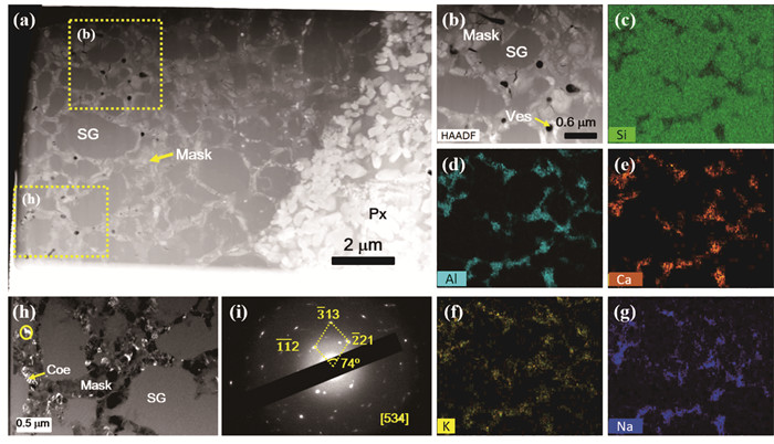
图 14 Ⅰ型柯石英的TEM分析
a, b. NWA 8657火星陨石FIB薄片的HAADF图像;c, g. 元素Si,Al,Ca,K和Na的EDXS分布图;h. 图a局部区域的TEM暗场像;i. 柯石英的选区电子衍射图谱(来图h圆圈处).SG.二氧化硅玻璃;Mask. 熔长石;Px. 辉石;Coe. 柯石英;Ves. 气孔.图据Hu et al.(2020)
Fig. 14. TEM analysis of Type Ⅰ coesite
表 1 透射电子显微镜的基本功能和相关技术
Table 1. Function and techniques of transmission electron microscope
分析功能 相关技术 获得信息 形貌像 BF-TEM、DF-TEM、SE 形貌、位错线和孪晶板条等 晶体结构与缺陷 SAED、CBED、NED、HRTEM、ACOM 晶体类型、晶粒取向、位错、孪晶、层错和界面结构等 原子成像 STEM-HAADF、ABF、DPC-STEM、iDPC-STEM 轻、重原子占位与分布、原子偏聚、原子电场/磁场分布 化学分析 (S)TEM-EDXS、STEM-EELS、EF-TEM 成分、原子尺度元素和空位偏析与分布、价态、键合成像等 磁结构表征 EH、Lorentz
TEM、EMCD磁场(电场)分布、磁交互作用、磁畴分布、磁化强度、轨道磁矩和自旋磁矩等 三维重构像 (S)TEM-3D ET、STEM-3D EDXS、AET 三维空间形态、结构和成分分布 原位和冷冻分析 In-situ TEM,cryo-TEM 实时、动态的形貌,成分和结构表征,特殊条件下(冷冻)电镜分析 注:BF-TEM. 明场像;DF-TEM. 暗场像;SE. 二次电子像;SAED. 选区电子衍射;CBED.会聚束电子衍射;NED. 纳米电子衍射;HRTEM. 高分辨透射电子显微像;TEM. 透射电子显微术(镜);ACOM. 自动晶体取向成像术;STEM. 扫描透射电子显微术(镜);STEM-HAADF. 高角环形暗场像;ABF. 环形明场像;DPC-STEM. 差分相位衬度成像;iDPC-STEM. 积分微分相位衬度成像;EDXS. X射线能量色散谱;EELS. 电子能量损失谱;EF-TEM. 能量过滤透射电子显微术;EH. 电子全息术;Lorentz TEM. 洛伦兹电子显微术;EMCD. 电子磁手性二向色性技术;3D ET. 电子三维重构技术;STEM-3D EDXS. X射线能量色散谱三维重构技术;AET. 原子电子三维重构技术;In-situ TEM. 原位透射电镜显微术;cryo-TEM. 冷冻透射电镜显微术.  下载: 导出CSV
表 2 TEM铜载网碳膜类型参数及其适用的纳米材料
Table 2. Types and applicability of carbon film coated TEM copper grid for nanomaterials
类别 特征 膜厚(nm) 衬度 适用样品 参考文献 纯碳膜 铜网和碳膜组成 15~30 一般 在有机溶剂或高温处理的材料(≥10 nm) Lu et al., 2013 微栅 有微孔的碳支持膜 15~20 优异 管状、棒状、纳米团聚物等 Tian et al., 2018 碳支持膜 铜网,方华膜和碳膜叠加 7~10 较好 粒径≥10 nm的纳米材料 Sun et al., 2019 超薄碳膜 在微栅上覆盖薄碳膜而成 3~5 优异 分散性好,粒径 < 10 nm的样品 Cai et al., 2019 双联网碳支持膜 2个碳支持膜相连,可折叠 7~10 一般 磁性纳米材料和矿物 He and Pan, 2020
下载: 导出CSV
-
Ahrenkiel, S.P., Yu, P.R., Murphy, J.E., et al., 2008. Nanoparticle Shape and Configuration Analysis by Transmission Electron Tomography. Journal of Microscopy, 230(3): 382-387. https://doi.org/10.1111/j.1365-2818.2008.01996.x Aitkaliyeva, A., Madden, J.W., Miller, B.D., et al., 2015. Comparison of Preparation Techniques for Nuclear Materials for Transmission Electron Microscopy (TEM). Journal of Nuclear Materials, 459: 241-246. https://doi.org/10.1016/j.jnucmat.2015.01.042 Allen, J.E., Hemesath, E.R., Perea, D.E., et al., 2008. High-Resolution Detection of Au Catalyst Atoms in Si Nanowires. Nature Nanotechnology, 3(3): 168-173. https://doi.org/10.1038/nnano.2008.5 Allen, L.J., D'Alfonso, A.J., Freitag, B., et al., 2012. Chemical Mapping at Atomic Resolution Using Energy-Dispersive X-Ray Spectroscopy. MRS Bulletin, 37(1): 47-52. https://doi.org/10.1557/mrs.2011.331 Ananth, M., Stern, L., Ferranti, D., et al., 2011. Creating Nanohole Arrays with the Helium Ion Microscope. Proceedings of SPIE, 8036: 80360M. https://doi.org/10.1117/12.887497 Asami, M., Suzuki, K., Grew, E.S., 2005. Monazite and Zircon Dating by the Chemical Th-U-Total Pb Isochron Method (ChIME) from Alasheyev Bight to the Sør Rondane Mountains, East Antarctica: A Reconnaissance Study of the Mozambique Suture in East Queen Maud Land. The Journal of Geology, 113(1): 59-82. https://doi.org/10.1086/425969 Bakken, B.M., Hochella, M.F., Marshall, A.F., et al., 1989. High-Resolution Microscopy of Gold in Unoxidized Ore from the Carlin Mine, Nevada. Economic Geology, 84(1): 171-179. https://doi.org/10.2113/gsecongeo.84.1.171 Benzerara, K., Bernard, S., Miot, J., 2019. Mineralogical Identification of Traces of Life. Springer International Publishing, New York, 123-144. Benzerara, K., Menguy, N., Lo'pez-Garcı'a, P., et al., 2006. Nanoscale Detection of Organic Signatures in Carbonate Microbialites. Proceedings of the National Academy of Sciences of the United States of America, 103(25): 9440-9445. https://doi.org/10.1073/pnas.0603255103 Benzerara, K., Menguy, N., Obst, M., et al., 2011. Study of the Crystallographic Architecture of Corals at the Nanoscale by Scanning Transmission X-Ray Microscopy and Transmission Electron Microscopy. Ultramicroscopy, 111(8): 1268-1275. https://doi.org/10.1016/j.ultramic.2011.03.023 Binig, G., Gerber, Ch., Stoll, E., et al., 1987. Atomic Resolution with Atomic Force Microscope. Surface Science Letters, 180-190: A390. https://doi.org/10.1016/0167-2584(87)90418-x Binnig, G., Rohrer, H., 1985. The Scanning Tunneling Microscope. Scientific American, 253(2): 50-56. https://doi.org/10.1038/scientificamerican0885-50 Bleloch, A., Lupini, A., 2004. Imaging at the Picoscale. Materials Today, 7(12): 42-48. https://doi.org/10.1016/S1369-7021(04)00570-x Boersch, H., 1946. Über die Möglichkeit der Abbildung von Atomen im Elektronenmikroskop. I. Monatshefte Für Chemie, 76(2): 86-92 (in German). doi: 10.1007/BF00898735 Brasier, M.D., Antcliffe, J., Saunders, M., et al., 2015. Changing the Picture of Earth's Earliest Fossils (3.5-1.9 Ga) with New Approaches and New Discoveries. PNAS, 112(16): 4859-4864. https://doi.org/10.1073/pnas.1405338111 Brasier, M.D., Wacey, D., 2012. Fossils and Astrobiology: New Protocols for Cell Evolution in Deep Time. International Journal of Astrobiology, 11(4): 217-228. doi: 10.1017/S1473550412000298 Broglie, L.D., 1924. Recherches Sur la Théorie Des Quanta. Annales de Physique, 10(Ⅲ): 22-128 (in French). Browning, N.D., Chisholm, M.F., Pennycook, S.J., 1993. Atomic-Resolution Chemical Analysis Using a Scanning Transmission Electron Microscope. Nature, 366(6451): 143-146. doi: 10.1038/366143a0 Bryson, J.F.J., Herrero-Albillos, J., Kronast, F., et al., 2014. Nanopaleomagnetism of Meteoritic Fe-Ni Studied Using X-Ray Photoemission Electron Microscopy. Earth and Planetary Science Letters, 396: 125-133. https://doi.org/10.1016/j.epsl.2014.04.016 Busch, H., 1927. Experimentelle Untersuchungen über Die Kohlensäurewirkung Auf Die Blutgefässe. Archiv für Elektrotechnik, 18(6): 583-594 (in German). doi: 10.1007/BF01656203 Buxton, B.F., Eades, J.A., Steeds, J.W., et al., 1976. The Symmetry of Electron Diffraction Zone Axis Patterns. Philosophical Transactions of the Royal Society A, 281: 181-184. Cai, Y., Wang, Y., Xu, H., et al., 2019. Positive Magnetic Resonance Angiography Using Ultrafine Ferritin-Based Iron Oxide Nanoparticles. Nanoscale, 11(6): 2644-2654. https://doi.org/10.1039/c8nr06812g Carter, C. B., Donald, A. M., Sass, S. L., 1980. The Study of Grain Boundary Thickness Using Electron Diffraction Techniques. Philosophical Magazine A, 41(4): 467-475. https://doi.org/10.1080/01418618008239326 Casey, J.D., Phaneuf, M., Chandler, C., et al., 2002. Copper Device Editing: Strategy for Focused Ion Beam Milling of Copper. Journal of Vacuum Science & Technology B, 20(6): 2682-2685. Cavosie, A.J., Wilde, S.A., Liu, D., et al., 2004. Internal Zoning and U-Th-Pb Chemistry of Jack Hills Detrital Zircons: A Mineral Record of Early Archean to Mesoproterozoic (4 348-1 576 Ma) Magmatism. Precambrian Research, 135(4): 251-279. https://doi.org/10.1016/j.precamres.2004.09.001 Chang, S.B.R., Kirschvink, J.L., 1989. Magnetofossils, the Magnetization of Sediments, and the Evolution of Magnetite Biomineralization. Annual Review of Earth and Planetary Sciences, 17(1): 169-195. https://doi.org/10.1146/annurev.ea.17.050189.001125 Chen, T.H., Chen, J., Ji, J.F., et al., 2005. Nanometer-Scale Investigation on the Loess of Luochuan: Nano-Rod Calcite. Geological Review, 51(6): 713-718, i0005-i0006(in Chinese with English abstract). Chen, T. H., Xu, H. F., Ji, J. F., et al., 2003. Formation Mechanism of Ferromagnetic Minerals in Loess of China: TEM Investigation. Chinese Science Bulletin, 48(17): 1883-1889(in Chinese). doi: 10.1360/csb2003-48-17-1883 Chen, X.F., Wang, Z.C., Zhong, X.Y., 2018. Developments of Energy-Filtered Transmission Electron Microscopy. Journal of Chinese Electron Microscopy Society, 37(5): 540-548 (in Chinese with English abstract). Chuvilin, A., Kaiser, U., 2005. On the Peculiarities of CBED Pattern Formation Revealed by Multislice Simulation. Ultramicroscopy, 104(1): 73-82. https://doi.org/10.1016/j.ultramic.2005.03.003 Cocherie, A., Legendre, O., Peucat, J.J., et al., 1998. Geochronology of Polygenetic Monazites Constrained by In Situ Electron Microprobe Th-U-Total Lead Determination Implications for Lead Behaviour in Monazite. Geochimica et Cosmochimica Acta, 62(14): 2475-2497. https://doi.org/10.1016/s0016-7037(98)00171-9 Cockayne, D. J. H., Parsons, J. R., Hoelke, C. W., 1971. A Study of the Relationship between Lattice Fringes and Lattice Planes in Electron Microscope Images of Crystals Containing Defects. The Philosophical Magazine: A Journal of Theoretical Experimental and Applied Physics, 24(187): 139-153. https://doi.org/10.1080/14786437108216429 Cockayne, D., McKenzie, D., Muller, D., 1991. Electron Diffraction of Amorphous Thin Films Using Peels. Microscopy Microanalysis Microstructures, 2(2-3): 359-366. https://doi.org/10.1051/mmm:0199100202-3035900 Crewe, A.V., Wall, J., Welter, L.M., 1968. A High-Resolution Scanning Transmission Electron Microscope. Journal of Applied Physics, 39(13): 5861-5868. https://doi.org/10.1126/science.6867711 Cui, J. P., Hao, Y. L., Li, S. J., et al., 2009. Reversible Movement of Homogenously Nucleated Dislocations in a Beta-Titanium Alloy. Physical Review Letters, 102(4): 045503. https://doi.org/10.1103/physrevlett.102.045503 D'Alfonso, A.J., Freitag, B., Klenov, D., et al., 2010. Atomic-Resolution Chemical Mapping Using Energy-Dispersive X-Ray Spectroscopy. Physical Review B, 81(10). https://doi.org/10.1103/PhysRevB.81.100101 Dahmen, U., Erni, R., Radmilovic, V., et al., 2009. Background, Status and Future of the Transmission Electron Aberration-Corrected Microscope Project. Philosophical Transactions Series A, Mathematical, Physical, and Engineering Sciences, 367(1903): 3795-3808. https://doi.org/10.1098/rsta.2009.0094 Davisson, C., Germer, L.H., 1927. Diffraction of Electrons by a Crystal of Nickel. Physical Review, 30(6): 705-740. https://doi.org/10.1103/PhysRev.30.705 De Rosier, D. J., Klug, A., 1968. Reconstruction of Three Dimensional Structures from Electron Micrographs. Nature, 217(5124): 130-134. https://doi.org/10.1038/217130a0 Dodd, M.S., Papineau, D., Grenne T., et al., 2017. Evidence for Early Life in Earth's Oldest Hydrothermal Vent Precipitates. Nature, 543(7643): 60-64. doi: 10.1038/nature21377 Du, H., Che, R., Kong, L., et al., 2015. Edge-Mediated Skyrmion Chain and Its Collective Dynamics in a Confined Geometry. Nature Communications, 6(1): 8504. https://doi.org/10.1038/ncomms9504 Du, H.J., 2005. The Preparation of TEM Specimen for the Nanomaterial Detection. Physical Testing and Chemical Analysis Part A (Physical Testing), 41(9): 463-466, 474(in Chinese with English abstract). Egerton, R.F., Malac, M., 2005. EELS in the TEM. Journal of Electron Spectroscopy and Related Phenomena, 143(2-3): 43-50. https://doi.org/10.1016/j.elspec.2003.12.009 Eun, J., 1991. Energy Dispersive Spectroscopy X-Ray Microanalysis. Journal of the Microelectronic Engineering Conference, 5(1): 31-38. Evans, M.E., Heller, F., 2001. Magnetism of Loess/Palaeosol Sequences: Recent Developments. Earth-Science Reviews, 54(1-3): 129-144. https://doi.org/10.1016/S0012-8252(01)00044-7 Fougerouse, D., Reddy, S.M., Saxey, D.W., et al., 2018. Nanoscale Distribution of Pb in Monazite Revealed by Atom Probe Microscopy. Chemical Geology, 479(1-3): 251-258. https://doi.org/10.1016/s0012-8252(01)00044-7 Fu, Q.Q., Shan, Z.W., 2016. FIB-SEM Dual-Beam System and Its Partial Applications. Journal of Chinese Electron Microscopy Society, 35(1): 81-89 (in Chinese with English abstract). Gao, P., Kumamoto, A., Ishikawa, R., et al., 2018. Picometer-Scale Atom Position Analysis in Annular Bright-Field STEM Imaging. Ultramicroscopy, 184(Pt A): 177-187. https://doi.org/10.1016/j.ultramic.2017.09.001 Giannuzzi, L. A., Drown, J. L., Brown, S. R., et al., 1998. Applications of the FIB Lift-out Technique for TEM Specimen Preparation. Microscopy Research and Technique, 41(4): 285-290. https://doi.org/10.1002/(sici)1097-0029(19980515)41:4<285:aid-jemt1>3.0.co;2-q doi: 10.1002/(sici)1097-0029(19980515)41:4<285:aid-jemt1>3.0.co;2-q Giannuzzia, L.A., Stevie, F.A., 1999. A Review of Focused Ion Beam Milling Techniques for TEM Specimen Preparation. Micron, 30(3): 197-204. https://doi.org/10.1016/S0968-4328(99)00005-0 Goncalves, P., Christian, N., Montel, J.M., 2004. Petrology and In Situ U-Th-Pb Monazite Geochronology of Ultrahigh-Temperature Metamorphism from the Andriamena Mafic Unit, North-Central Madagascar: Significance of a Petrographical P-T Path in a Polymetamorphic Context. Journal of Petrology, 45(10): 1923-1957. https://doi.org/10.1093/petrology/egh041 Gu, L., Zhu, C. B., Li, H., et al., 2011. Direct Observation of Lithium Staging in Partially Delithiated LiFePO4 at Atomic Resolution. Journal of the American Chemical Society, 133(13): 4661-4663. https://doi.org/10.1021/ja109412x Gu, L.X., Li, J.H., 2020. Focused Ion Beam (FIB) Technology and Its Applications for Earth and Planetary Sciences. Bulletin of Mineralogy, Petrology and Geochemistry, 39: 1-22 (in Chinese with English abstract). Gu, L.X., Zhang, B., Hu, S., et al., 2018. The Discovery of Silicon Oxide Nanoparticles in Space-Weathered of Apollo 15 Lunar Soil Grains. Icarus, 303: 47-52. https://doi.org/10.1016/j.icarus.2017.12.028 Guo, Z., Li, Y., Liu, S., et al., 2020. Discovery of Nanophase Iron Particles and High Pressure Clinoenstatite in a Heavily Shocked Ordinary Chondrite: Implications for the Decomposition of Pyroxene. Geochimica et Cosmochimica Acta, 272: 276-286. https://doi.org/10.1016/j.gca.2019.10.036 Hagemann, P., Thompson, M.N., 1983. Analytical Capabilities of Transmission Electron Microscope (TEM) Systems. Microscopy: Techniques and Capabilities, 368: 29-34. https://doi.org/10.1117/12.934321 Hagler, H.K., 2007. Ultramicrotomy for Biological Electron Microscopy. Electron Microscopy, 369: 67-96. https://doi.org/10.1007/978-1-59745-294-6_5 Haguenau, F., Hawkes, P. W., Hutchison, J. L., et al., 2003. Key Events in the History of Electron Microscopy. Microscopy and Microanalysis, 9(2): 96-138. https://doi.org/10.1017/s1431927603030113 Haider, M., Rose, H., Uhlemann, S., et al., 1998. A Spherical-Aberration-Corrected 200 kV Transmission Electron Microscope. Ultramicroscopy, 75: 53-60. https://doi.org/10.1016/s0304-3991(98)00048-5 Haider, M., Uhlemann, S., Zach, J., 2000. Upper Limits for the Residual Aberrations of a High-Resolution Aberration-Corrected STEM. Ultramicroscopy, 81(3/4): 163-175. https://doi.org/10.1016/s0304-3991(99)00194-1 Han, W., Xiao, S.Q., 2013. Focused Ion Beam (FIB) and Its Applications. Materials China, 32(12): 716-727 (in Chinese with English abstract). Han, X.D., Zhang, Z., 2010. In Situ Mechanical Experiments at Atomic Lattice Resolution. Journal of Chinese Electron Microscopy Society, 29(3): 191-212 (in Chinese with English abstract). Hankamer, B., Glaeser, R., Stahlberg, H., 2007. Electron Crystallography of Membrane Proteins. Journal of Structural Biology, 160(3): 263-264. https://doi.org/10.1016/j.jsb.2007.11.001 Harte, B., 1994. Lower Mantle Mineral Associations Preserved in Diamonds. Mineralogical Magazine, 58A(1): 384-385. doi: 10.1180/minmag.1994.58A.1.201 Hayman, P.C., Kopylova, M.G., Kaminsky, F.V., 2005. Lower Mantle Diamonds from Rio Soriso (Juina Area, Mato Grosso, Brazil). Contributions to Mineralogy and Petrology, 149(4): 430-445. https://doi.org/10.1007/s00410-005-0657-8 He, H.P., Zhu, J.X., Chen, M., et al., 2020. Progresses in Researches on Mineral Structure and Mineral Physics (2011—2020). Bulletin of Mineralogy, Petrology and Geochemistry, 39(4): 697-713 (in Chinese with English abstract). He, K., Pan, Y., 2020. Magnetofossil Abundance and Diversity as Paleoenvironmental Proxies: A Case Study from Southwest Iberian Margin Sediments. Geophysical Research Letters, 47(8). https://doi.org/10.1029/2020gl087165 Heaney, P.J., Vicenzi, E.P., Giannuzzi, L.A., et al., 2001. Focused Ion Beam Milling: A Method of Site-Specific Sample Extraction for Microanalysis of Earth and Planetary Materials. American Mineralogist, 86(9): 1094-1099. https://doi.org/10.2138/am-2001-8-917 Heller, F., Liu, T.S., 1982. Magnetostratigraphical Dating of Loess Deposits in China. Nature, 300(5891): 431-433. doi: 10.1038/300431a0 Henderson, R., 1989. A Model for the Structure of Bacteriorhodopsin Based on High-Resolution Cryoelectron Microscopy. Ultramicroscopy, 31(4): 467. https://doi.org/10.1016/0304-3991(89)90371-9 Heslop, D., 2015. Numerical Strategies for Magnetic Mineral Unmixing. Earth-Science Reviews, 150: 256-284. https://doi.org/10.1016/j.earscirev.2015.07.007 Heslop, D., 2009. On the Statistical Analysis of the Rock Magnetic S-Ratio. Geophysical Journal International, 178(1): 159-161. https://doi.org/10.1111/j.1365-246x.2009.04175.x Hillebrand, R., Pippel, E., Hesse, D., et al., 2011. A Study of Intermixing in Perovskite Superlattices by Simulation-Supported Cs-Corrected HAADF-STEM. Physica Status Solidi (A), 208(9): 2144-2149. https://doi.org/10.1016/j.earscirev.2015.07.007 Hochella, Jr., M.F., 2002a. Nanoscience and Technology: The Next Revolution in the Earth Sciences. Earth and Planetary Science Letters, 203(2): 593-605. https://doi.org/10.1016/s0012-821x(02)00818-x Hochella, Jr., M.F., 2002b. There's Plenty of Room at the Bottom Nanoscience in Geochemistry. Geochimica et Cosmochimica Acta, 66(5): 735-743. https://doi.org/10.1016/s0016-7037(01)00868-7 Hochella, Jr., M.F., Lower, S.K., Maurice, P.A., et al., 2008. Nanominerals, Mineral Nanoparticles, and Earth Systems. Science, 319(5870): 1631-1635. https://doi.org/10.1126/science.1141134 Hochella, Jr., M.F., Mogk, D.W., Ranville, J., et al., 2019. Natural, Incidental, and Engineered Nanomaterials and Their Impacts on the Earth System. Science, 363(6434): eaau8299. https://doi.org/10.1126/science.aau8299 Hochella, Jr., M. F., Spencer, M. G., et al., 2015. Nanotechnology: Nature's Gift or Scientists' Brainchild? Environmental Science: Nano, 2(2): 114-119. https://doi.org/10.1039/c4en00145a Hoppe, P., Cohen, S., Meibom, A., 2013. NanoSIMS: Technical Aspects and Applications in Cosmochemistry and Biological Geochemistry. Geostandards and Geoanalytical Research, 37(2): 111-154. doi: 10.1111/j.1751-908X.2013.00239.x Hrncir, T., Hladik, L., Zadrazil, M., 2013. Fast 3D Tomography at Package Level by Using Xe Plasma Focused Ion Beam. Proceedings of the 20th IEEE International Symposium on the Physical and Failure Analysis of Integrated Circuits (IPFA), Suzhou, 112-115. Hu, S., Li, Y., Gu, L.X., et al., 2020. Discovery of Coesite from the Martian Shergottite Northwest Africa 8657. Geochimica et Cosmochimica Acta, 286: 404-417. https://doi.org/10.1016/j.gca.2020.07.021 Huang, L.Y., Wang, K.D., Jin, H.M., et al., 1981. The Design and Adjustment of a DX-4 Transmission Electron Microscope. Chinese Journal of Scientific Instrument, 2(2): 1-9 (in Chinese with English abstract). Huang, Y.Z., Lozano-Perez, S., Langford, R.M., et al., 2002. Preparation of Transmission Electron Microscopy Cross-Section Specimens of Crack Tips Using Focused Ion Beam Milling. Journal of Microscopy, 207(2): 129-136. https://doi.org/10.1116/1.1378072 Huang, Z., Fryer, J.R., Park, C., et al., 1996. Transmission Electron Microscopy and Energy Dispersive X-Ray Spectroscopy Studies of Pt-Re /γ-Al2O3 Catalysts. Journal of Catalysis, 148(2): 478-492. https://doi.org/10.1006/jcat.1994.1234 Hughes, A., 1955. Studies in the History of Microscopy. Journal of Microscopy, 75(1): 1-22. https://doi.org/10.1111/j.1365-2818.1955.tb00403.x Iijima, S., 1971. High-Resolution Electron Microscopy of Crystal Lattice of Titanium-Niobium Oxide. Journal of Applied Physics, 42(13): 5891-5893. https://doi.org/10.1063/1.1660042 Ishikawa, R., Findlay, S.D., Seki, T., et al., 2018. Direct Electric Field Imaging of Graphene Defects. Nature Communications, 9(1): 3878. https://doi.org/10.1038/s41467-018-06387-8 Ishikawa, R., Okunishi, E., Sawada, H., et al., 2011. Direct Imaging of Hydrogen-Atom Columns in a Crystal by Annular Bright-Field Electron Microscopy. Nature Materials, 10(4): 278-281. doi: 10.1038/nmat2957 James, E.M., Browning, N.D., 1999. Practical Aspects of Atomic Resolution Imaging and Analysis in STEM. Ultramicroscopy, 78(1-4): 125-139. https://doi.org/10.1016/s0304-3991(99)00018-2 Jia, Z.H., Ding, L.P., Chen, H.W., 2015. The Principle and Applications of High-Resolution Scanning Electron Microscopy. Physics, 44(7): 446-452 (in Chinese with English abstract). Jiang, Y., Chen, Z., Han, Y., et al., 2018. Electron Ptychography of 2D Materials to Deep Sub-ÅNGSTRÖM Resolution. Nature, 559(7714): 343-349. https://doi.org/10.1038/s41586-018-0298-5 Jongmans, A.G., van Oort, F., Denaix, L., et al., 1999. Mineral Micro- and Nano-Variability Revealed by Combined Micromorphology and In Situ Submicroscopy. Catena, 35(2-4): 259-279. https://doi.org/10.1016/s0341-8162(98)00104-0 Ju, Y.W., Huang, C., Sun, Y., et al., 2018. Nanogeoscience: Connotation and Significance. Earth Science, 43(5): 1367-1383 (in Chinese with English abstract). Kaminsky, F.V., Wirth, R., 2011. Iron Carbide Inclusions in Lower-Mantle Diamond from Juina, Brazil. The Canadian Mineralogist, 49(2): 555-572. https://doi.org/10.3749/canmin.49.2.555 Kaminsky, F.V., Wirth, R., Schreiber, A., 2013. Carbonatitic Inclusions in Deep Mantle Diamond from Juina, Brazil: New Minerals in the Carbonate-Halide Association. The Canadian Mineralogist, 51(5): 669-688. https://doi.org/10.3749/canmin.51.5.669 Kauschi, V.G.A., Pfankuch, E., Ruska, H., 1939. Die Sichtbarmachung Yon Pflanzlichem Virus Im Obermikroskop. Naturwissenschaften, 27(18): 292-299 (in German). doi: 10.1007/BF01493353 Kesson, S.E., Fitz Gerald, J.D., Shelley, J.M., 1998. Mineralogy and Dynamics of a Pyrolite Lower Mantle. Nature, 393(6682): 252-255. doi: 10.1038/30466 Kotula, P.G., Keenan, M.R., 2006. Application of Multivariate Statistical Analysis to STEM X-Ray Spectral Images: Interfacial Analysis in Microelectronics. Microscopy and Microanalysis, 12(6): 538-544. https://doi.org/10.1017/s1431927606060636 Kral, M.V., Spanos, G., 1999. Three-Dimensional Analysis of Proeutectoid Cementite Precipitates. Acta Materialia, 47(2): 711-724. https://doi.org/10.1016/S1359-6454(98)00321-8 Krivanek, O.L., Dellby, N., Lupini, A. R., 1999. Towards Sub-Angstrom Electron Beams. Ultramicroscopy, 78(1-4): 1-11. doi: 10.1016/S0304-3991(99)00013-3 Krivanek, O.L., Gubbens, A.J., Dellby, N., 1991. Developments in EELS Instrumentation for Spectroscopy and Imaging. Microscopy Microanalysis Microstructures, 2(2-3): 315-332. https://doi.org/10.1051/mmm:0199100202-3031500 Kübel, C., Voigt, A., Schoenmakers, R., et al., 2005. Recent Advances in Electron Tomography: TEM and HAADF-STEM Tomography for Materials Science and Semiconductor Applications. Microscopy and Microanalysis, 11(5): 378-400. https://doi.org/10.1017/s1431927605050361 Kühlbrandt, W., 2014. Cryo-Em Enters a New Era. Elife, 3: e03678. https://doi.org/10.7554/eLife.03678 Kusiak, M.A., Dunkley, D.J., Wirth, R., et al., 2015. Metallic Lead Nanospheres Discovered in Ancient Zircons. Proceedings of the National Academy of Sciences of the United States of America, 112(16): 4958-4963. doi: 10.1073/pnas.1415264112 Lazic, I., Bosch, E.G.T., Lazar, S., 2016. Phase Contrast Stem for Thin Samples: Integrated Differential Phase Contrast. Ultramicroscopy, 160: 265-280. https://doi.org/10.1016/j.ultramic.2015.10.011 Le Guillou, C., Bernard, S., Brearley, A.J., et al., 2014. Evolution of Organic Matter in Orgueil, Murchison and Renazzo during Parent Body Aqueous Alteration: In Situ Investigations. Geochimica et Cosmochimica Acta, 131: 368-392. https://doi.org/10.1016/j.gca.2013.11.020 Leapman, R.D., Hunt, J.A., 1991. Comparison of Detection Limits for EELS and EDXS. Microscopy Microanalysis Microstructures, 2(2-3): 231-244. https://doi.org/10.1051/mmm:0199100202-3023100 Lepinay, K., Lorut, F., Pofelski, A., et al., 2013. Defect Analysis of a Silicon Nanowire Transistor by X-Ray Energy Dispersive Spectroscopy Technique in a STEM: 2D Mappings and Tomography. Journal of Physics: Conference Series, 471: 012027. doi: 10.1088/1742-6596/471/1/012027 Lepot, K., Benzerara, K., Brown, G.E., et al., 2008. Microbially Influenced Formation of 2, 724-Million-Year-Old Stromatolites. Nature Geoscience, 1(2): 118-121. https://doi.org/10.1038/ngeo107 Li, C., Yang, G., 2014. The Principle and Applications of STEM and EELS. Physics, 43(9): 597-605 (in Chinese with English abstract). Li, D.X., 2004a. Progress of Transmission Electron Microscopy I Development of Transmission Electron Microscope and Related Equipments. Journal of Chinese Electron Microscopy Society, 23(3): 269-277 (in Chinese with English abstract). Li, D.X., 2004b. Progress of Transmission Electron Microscopy Ⅱ Z-Contrast Imaging, Sub-Angstrom Transmission Electron Microscopy, Aberration-Corrected Transmission Electron Microscopy. Journal of Chinese Electron Microscopy Society, 23(3): 278-292(in Chinese with English abstract). Li, J.H., Benzerara, K., Bernard, S., et al., 2013a. The Link between Biomineralization and Fossilization of Bacteria: Insights from Field and Experimental Studies. Chemical Geology, 359: 49-69. https://doi.org/10.1016/j.chemgeo.2013.09.013 Li, J.H., Ge, K.P., Pan, Y.X., et al., 2013b. A Strong Angular Dependence of Magnetic Properties of Magnetosome Chains: Implications for Rock Magnetism and Paleomagnetism. Geochemistry, Geophysics, Geosystems, 14(10): 3887-3907. doi: 10.1002/ggge.20228 Li, J.H., Menguy, N., Gatel, C., et al., 2015. Crystal Growth of Bullet-Shaped Magnetite in Magnetotactic Bacteria of the Nitrospirae Phylum. Journal of the Royal Society Interface, 12(103): 20141288. https://doi.org/10.1002/9783527808465.emc2016.6869 Li, J.H., Pan, Y.X., 2015. Applications of Transmission Electron Microscopy in the Earth Sciences. Scientia Sinica Terrae, 45(9): 1359-1382 (in Chinese). doi: 10.1360/zd2015-45-9-1359 Li, J.H., Zhang, H., Liu, P.Y., et al., 2019. Phylogenetic and Structural Identification of a Novel Magnetotactic Deltaproteobacteria Strain, Wyhr-1, from a Freshwater Lake. Applied and Environmental Microbiology, 85: e00731-00719. https://doi.org/10.1128/aem.00731-19 Li, J.H., Zhang, H., Menguy, N., et al., 2017a. Single-Cell Resolution of Uncultured Magnetotactic Bacteria via Fluorescence-Coupled Electron Microscopy. Applied and Environmental Microbiology, 83(12): e00409-00417. https://doi.org/10.1128/AEM.00409-17 Li, Y.L., Konhauser, K.O., Zhai, M.G., 2017b. The Formation of Magnetite in the Early Archean Oceans. Earth and Planetary Science Letters, 466: 103-114. https://doi.org/10.1016/j.epsl.2017.03.013 Li, Z.A., Zheng, F.S., Tavabi, A.H., et al., 2017c. Magnetic Skyrmion Formation at Lattice Defects and Grain Boundaries Studied by Quantitative Off-Axis Electron Holography. Nano Letters, 17(3): 1395-1401. https://doi.org/10.1021/acs.nanolett.6b04280 Li, J.H., Liu, P.Y., Wang, J., et al., 2020a. Magnetotaxis as an Adaptation to Enable Bacterial Shuttling of Microbial Sulfur and Sulfur Cycling across Aquatic Oxic-Anoxic Interfaces. Journal of Geophysical Research: Biogeosciences, 125(12): e2020JG006012. https://doi.org/10.1029/2020jg006012 Li, J.H., Liu, Y., Liu, S.C., et al., 2020b. Classification of a Complexly Mixed Magnetic Mineral Assemblage in Pacific Ocean Surface Sediment by Electron Microscopy and Supervised Magnetic Unmixing. Frontiers in Earth Science, 8: 609058. https://doi.org/10.3389/feart.2020.609058 Li, J.H., Menguy, N., Leroy, E., et al., 2020c. Biomineralization and Magnetism of Uncultured Magnetotactic Coccus Strain Thc-1 with Non-Chained Magnetosomal Magnetite Nanoparticles. Journal of Geophysical Research: Solid Earth, 125(12): e2020JB020853. https://doi.org/10.1029/2020jb020853 Li, J.H., Margaret, Oliver, I., Cam, N., et al., 2016. Biomineralization Patterns of Intracellular Carbonatogenesis in Cyanobacteria: Molecular Hypotheses. Minerals, 6(1): 10. https://doi.org/10.3390/min6010010 Li, J.H., Menguy, N., Roberts, A.P., et al., 2020d. Bullet-Shaped Magnetite Biomineralization within a Magnetotactic Deltaproteobacterium: Implications for Magnetofossil Identification. Journal of Geophysical Research: Biogeosciences, 125: e2020JG005680. https://doi.org/10.1029/2020jg005680 Li, J.H., Pan, Y.X., Liu, Q.S., et al., 2010. Biomineralization, Crystallography and Magnetic Properties of Bullet-Shaped Magnetite Magnetosomes in Giant Rod Magnetotactic Bacteria. Earth and Planetary Science Letters, 293(3-4): 368-376. https://doi.org/10.1016/j.epsl.2010.03.007 Li, J.H., Pei, R., Teng, F.F., et al., 2021. Micro-XRF Study of the Troodontid Dinosaur Jianianhualong Tengi Reveals New Biological and Taphonomical Signals. Atomic Spectroscopy, 41(6): 1-11. https://doi.org/10.1101/2020.09.07.285833 Li, Q.L., Yang, W., Liu, Y., et al., 2013. Ion Microprobe Microanalytical Techniques and Their Applications in Earth Sciences. Bulletin of Mineralogy, Petrology and Geochemistry, 32(3): 310-327 (in Chinese with English abstract). Liu, J., Cowley, J.M., 1993. High-Resolution Scanning Transmission Electron Microscopy. Ultramicroscopy, 52(3-4): 335-346. https://doi.org/10.1016/0304-3991(93)90044-x Liu, P.Y., Liu, Y., Zhao, X., et al., 2020. Diverse Phylogeny and Morphology of Magnetite Biomineralized by Magnetotactic Cocci. Environmental Microbiology, 23(2): 1115-1129. https://doi.org/10.1111/1462-2920.15254 Liu, Q.S., Roberts, A.P., Larrasoaña, J.C., et al., 2012. Environmental Magnetism: Principles and Applications. Reviews of Geophysics, 50(4): RG4002. https://doi.org/10.1029/2012RG000393 Longo, P., Thomas, P.J., Twesten, R.D., 2012. Atomic-Level EELS Mapping Using High-Energy Edges in DualeelsTM Mode. Microscopy Today, 20(4): 30-36. https://doi.org/10.1017/S1431927612003844 Lu, P., Boyle, T., Clark, B., et al., 2013. In Situ TEM Heating Study of Cu and Ag Nanoparticle Interaction. Microscopy and Microanalysis, 19(S2): 448-449. https://doi.org/10.1017/S1431927613004236 Lu, X., Zhao, L., He, X., et al., 2012. Lithium Storage in Li4Ti5O12 Spinel: The Full Static Picture from Electron Microscopy. Advanced Materials (Deerfield Beach, Fla), 24(24): 3233-3238. https://doi.org/10.1002/adma.201200450 Ma, Y.H., Cai, D.L., Li, Y.R., et al., 2016. The Influence of Straight Pore Blockage on the Selectivity of Methanol to Aromatics in Nanosized Zn/ZSM-5: An Atomic Cs-Corrected STEM Analysis Study. RSC Advances, 6(78): 74797-74801. https://doi.org/10.1039/c6ra19073a Maher, B.A., Thompson, R., 1995. Paleorainfall Reconstructions from Pedogenicmagnetic Susceptibility Variations in the Chinese Loess and Paleosols. Quaternary Research, 44(3): 383-391. https://doi.org/10.1006/qres.1995.1083 Malis, T. F., Steele, D., 1990. Ultramicrotomy for Materials Science. MRS Proceedings, 199: 3-42. doi: 10.1557/PROC-199-3 Malis, T., Cheng, S.C., Egerton, R.F., 1988. EELS Log-Ratio Technique for Specimen-Thickness Measurement in the TEM. Journal of Electron Microscopy Technique, 8(2): 193-200. https://doi.org/10.1002/jemt.1060080206 Mann, S., Frankel, R.B., Blakemore, R.P., 1984. Structure, Morphology and Crystal Growth of Bacterial Magnetite. Nature, 310(5976): 405-407. https://doi.org/10.1038/310405a0 Martin, L.C., Whelpton, R.V., Parnum, D.H., 1937. A New Electron Microscope. Journal of Scientific Instruments, 14(1): 14-24. doi: 10.1088/0950-7671/14/1/303 McCaffrey, J.P., Phaneuf, M.W., Madsen, L.D., 2001. Surface Damage Formation during Ion-Beam Thinning of Samples for Transmission Electron Microscopy. Ultramicroscopy, 87(3): 97-104. https://doi.org/10.1016/s0304-3991(00)00096-6 McVitie, S., Cushley, M., 2006. Quantitative Fresnel Lorentz Microscopy and the Transport of Intensity Equation. Ultramicroscopy, 106(4/5): 423-431. https://doi.org/10.1016/j.ultramic.2005.12.001 Meldrum, A., Boatner, L.A., Weber, W.J., et al., 1998. Radiation Damage in Zircon and Monazite. Geochimica et Cosmochimica Acta, 62(14): 2509-2520. https://doi.org/10.1016/s0016-7037(98)00174-4 Meldrum, F.C., Mann, S., Heywood, B.R., et al., 1993. Electron Microscopy Study of Magnetosomes in a Cultured Coccoid Magnetotactic Bacterium. Proceedings of the Royal Society B, 251(1332): 231-236. https://doi.org/10.1098/rspb.1993.0034 Miao, J.W., Ercius, P., Billinge, S.J.L., 2016. Atomic Electron Tomography: 3D Structures without Crystals. Science, 353(6306): aaf2157. https://doi.org/10.1126/science.aaf2157 Midgley, P. A., Dunin-Borkowski, R. E., 2009. Electron Tomography and Holography in Materials Science. Nature Materials, 8(4): 271-280. https://doi.org/10.1038/nmat2406 Mitsuishi, K., Hashimoto, A., Takeguchi, M., et al., 2010. Imaging Properties of Bright-Field and Annular-Dark-Field Scanning Confocal Electron Microscopy. Ultramicroscopy, 111(1): 20-26. https://doi.org/10.1016/j.ultramic.2011.10.004 Mook, H.W., Kruit, P., 2000. Construction and Characterization of the Fringe Field Monochromator for a Field Emission Gun. Ultramicroscopy, 81(3-4): 129-139. https://doi.org/10.1016/S0304-3991(99)00193-x Moorbath, S., 2005. Dating Earliest Life. Nature, 434: 155. doi: 10.1038/434155a Morishita, S., Ishikawa, R., Kohno, Y., et al., 2018. Attainment of 40.5 Pm Spatial Resolution Using 300 kV Scanning Transmission Electron Microscope Equipped with Fifth-Order Aberration Corrector. Microscopy, 67(1): 46-50. https://doi.org/10.1093/jmicro/dfx122 Muller, D.A., Kourkoutis, L.F., Murfitt, M., et al., 2008. Atomic-Scale Chemical Imaging of Composition and Bonding by Aberration-Corrected Microscopy. Science, 319(5866): 1073-1076. https://doi.org/10.1126/science.1148820 Muller, K., Krause, F.F., Beche, A., et al., 2014. Atomic Electric Fields Revealed by a Quantum Mechanical Approach to Electron Picodiffraction. Nature Communications, 5(1): 5653. doi: 10.1038/ncomms6653 Mulligan, S. K., Speir, J. A., Razinkov, I., et al., 2015. Multiplexed TEM Specimen Preparation and Analysis of Plasmonic Nanoparticles. Microscopy and Microanalysis, 21(4): 1017-1025. https://doi.org/10.1017/s1431927615014324 Nellist, P.D., 2012. Atomic Resolution Comes into Phase. Nature Physics, 8(8): 586-587. doi: 10.1038/nphys2357 Nepijko, S. A., Schönhense, G., 2009. Quantitative Lorentz Transmission Electron Microscopy of Structured Thin Permalloy Films. Applied Physics A, 96(3): 671-677. https://doi.org/10.1007/s00339-009-5131-4 Nie, J. F., Zhu, Y. M., Liu, J. Z., et al., 2013. Periodic Segregation of Solute Atoms in Fully Coherent Twin Boundaries. Science, 340(6135): 957-960. https://doi.org/10.1126/science.1229369 Noguchi, T., Kimura, M., Hashimoto, T., et al., 2014. Space Weathered Rims Found on the Surfaces of the Itokawa Dust Particles. Meteoritics & Planetary Science, 49(2): 188-214. https://doi.org/10.1111/maps.12111 Oatley, C.W., 1982. The Early History of the Scanning Electron Microscope. Journal of Applied Physics, 53(2): R1-R13. https://doi.org/10.1016/s1076-5670(04)33002-8 Obst, M., Gasser, P., Mavrocordatos, D., et al., 2005. TEM-Specimen Preparation of Cell/Mineral Interfaces by Focused Ion Beam Milling. American Mineralogist, 90(8-9): 1270-1277. https://doi.org/10.2138/am.2005.1743 O'Keefe, M.A., 2008. Seeing Atoms with Aberration-Corrected Sub-Angström Electron Microscopy. Ultramicroscopy, 108(3): 196-209. https://doi.org/10.1016/j.ultramic.2007.07.009 Okunishi, E., Sawada, H., Kondo, Y., et al., 2006. Atomic Resolution Elemental Map of EELS with a Cs Corrected STEM. Microscopy and Microanalysis, 12(S02): 1150-1151. https://doi.org/10.1017/s1431927606067481 Okunishi, E., Ishikawa, I., Sawada, H., et al., 2009. Visualization of Light Elements at Ultrahigh Resolution by Stem Annular Bright Field Microscopy. Microscopy and Microanalysis, 15(S2): 164-165. https://doi.org/10.1017/S1431927609093891 Otten, M.T., Coene, W.M.J., 1993. High-Resolution Imaging on a Field Emission TEM. Ultramicroscopy, 48(1-2): 77-91. doi: 10.1016/0304-3991(93)90173-U Oue, S., Nakano, H., Kuroda, R., et al., 2006. TEM-EDX Observations of the Microstructure of Electrodeposited Ni-Sn Alloys. Materials Transactions, 47(6): 1550-1554. https://doi.org/10.2320/jinstmet.70.804 Paasche, Ø., Løvlie, R., Dahl, S.O., et al., 2004. Bacterial Magnetite in Lake Sediments: Late Glacial to Holocene Climate and Sedimentary Changes in Northern Norway. Earth and Planetary Science Letters, 223(3-4): 319-333. https://doi.org/10.1016/j.epsl.2004.05.001 Palenik, C.S., Utsunomiya, S., Reich, M., et al., 2004. "Invisible" Gold Revealed: Direct Imaging of Gold Nanoparticles in a Carlin-Type Deposit. American Mineralogist, 89(10): 1359-1366. https://doi.org/10.2138/am-2004-1002 Pan, Y. X., Deng, C. L., Liu, Q. S., et al., 2004. Biomineralization and Magnetism of Bacterial Magnetosomes. Chinese Science Bulletin, 49(24): 2505-2510 (in Chinese). doi: 10.1360/csb2004-49-24-2505 Pan, Y.H., Zheng, W.X., Moyer, A. E., et al., 2016. Molecular Evidence of Keratin and Melanosomes in Feathers of the Early Cretaceous Bird Eoconfuciusornis. Proceedings of the National Academy of Sciences of the United States of America, 113(49): E7900-E7907. https://doi.org/10.1073/pnas.1617168113 Park, H.S., Murakami, Y., Yanagisawa, K., et al., 2012. Electron Holography Studies on Narrow Magnetic Domain Walls Observed in a Heusler Alloy Ni50Mn25Al12.5Ga12.5. Advanced Functional Materials, 22(16): 3434-3437. https://doi.org/10.1002/adfm.201103052 Pennycook, S.J., Boatner, L.A., 1988. Chemically Sensitive Structure-Imaging with a Scanning Transmission Electron Microscope. Nature, 336(6199): 565-567. doi: 10.1038/336565a0 Pennycook, S.J., Jesson, D., 1990. High-Resolution Incoherent Imaging of Crystals. Physical Review Letters, 64(8): 938-941. http://dx.doi.org/10.1103/PhysRevLett.64.938 Pennycook, S.J., Jesson, D.E., 1991. High-Resolution Z-Contrast Imaging of Crystals. Ultramicroscopy, 37(1-4): 14-38. doi: 10.1016/0304-3991(91)90004-P Pennycook, S.J., Jesson, D.E., 1992. Atomic Resolution Z-Contrast Imaging of Interfaces. Acta Metallurgica et Materialia, 40: S149-S159. http://dx.doi.org/10.1016/0956-7151(92)90275-J Pennycook, S.J., Varela, M., Hetherington, C.J.D., et al., 2006. Materials Advances through Aberration Corrected Electron Microscopy. MRS Bulletin, 31(1): 36-43. http://dx.doi.org/10.1557/mrs2006.4 Philippot, P., van Zuilen, M., Lepot, K., et al., 2007. Early Archaean Microorganisms Preferred Elemental Sulfur, Not Sulfate. Science, 317(5844): 1534-1537. doi: 10.1126/science.1145861 Phillipp, F., Höschen, R., Osaki, M., et al., 1994. New High-Voltage Atomic Resolution Microscope Approaching 1 Å Point Resolution Installed in Stuttgart. Ultramicroscopy, 56(1-3): 1-10. https://doi.org/10.1016/0304-3991(94)90141-4 Pidgeon, R. T., O'Neil, J. R., Silver, L. T., 1966. Uranium and Lead Isotopic Stability in a Metamict Zircon under Experimental Hydrothermal Conditions. Science, 154(3756): 1538-1540. https://doi.org/10.1126/science.154.3756.1538 Qin, S., Wang, R.C., 2004. Geometric Descriptions of Distorted Structures of ABX3 Type Perovskite and Application in Structural Prediction. Acta Geologica Sinica, 78(3): 345-351 (in Chinese with English abstract). Rauch, E.F., Véron, M., 2014. Automated Crystal Orientation and Phase Mapping in TEM. Materials Characterization, 98: 1-9. https://doi.org/10.1016/j.matchar.2014.08.010 Reich, M., Utsunomiya, S., Kesler, S.E., et al., 2006. Thermal Behavior of Metal Nanoparticles in Geologic Materials. Geology, 34(12): 1033. doi: 10.1130/G22829A.1 Reimer, L., Fromm, I., Hirsch, P., et al., 1992. Combination of EELS Modes and Electron Spectroscopic Imaging and Diffraction in an Energy-Filtering Electron Microscope. Ultramicroscopy, 46(1-4): 335-347. https://doi.org/10.1016/0304-3991(92)90023-d Reyntjens, S., Puers, R., 2001. A Review of Focused Ion Beam Applications in Microsystem Technology. Journal of Micromechanics and Microengineering, 11(4): 287-300. https://doi.org/10.1088/0960-1317/11/4/301 Ricolleau, C., Nelayah, J., Oikawa, T., et al., 2012. High Resolution Imaging and Spectroscopy Using Cs-Corrected TEM with Cold FEG JEM-ARM200F. JEOL News, 47(1): 2-8. Roberts, A.P., Hu, P.X., Harrison, R.J., et al., 2019. Domain State Diagnosis in Rock Magnetism: Evaluation of Potential Alternatives to the Day Diagram. Journal of Geophysical Research: Solid Earth, 124(6): 5286-5314. https://doi.org/10.1029/2018jb017049 Rose, H., 1990. Outline of a Spherically Corrected Semiaplanatic Medium-Voltage Transmission Electron Microscope. Optik, 85: 19-24. Rouvimov, S., Moeck, P., Häusler, I., et al., 2011. Automated Crystallite Orientation and Phase Mapping in a Transmission Electron Microscope. MRS Proceedings, 1318: 37-42. https://doi.org/10.1557/opl.2011.921 Rubino, S., Schattschneider, P., Stöger-Pollach, M., et al., 2007. EMCD Magnetic Chiral Dichroism in the Electron Microscope. MRS Proceedings, 1026: 1305. Ruprecht, J., Nield, J., 2001. Determining the Structure of Biological Macromolecules by Transmission Electron Microscopy, Single Particle Analysis and 3D Reconstruction. Progress in Biophysics and Molecular Biology, 75(3): 121-164. https://doi.org/10.1016/S0079-6107(01)00004-9 Ruska, E., 1934. Über Fortschritte Im Bau Und in Der Leistung des Magnetischen Elektronenmikroskops. Zeitschrift Für Physik, 87(9/10): 580-602 (in German). https://doi.org/10.1007/bf01333326 Sánchez-Santolino, G., Lugg, N. R., Seki, T., et al., 2018. Probing the Internal Atomic Charge Density Distributions in Real Space. ACS Nano, 12(9): 8875-8881. https://doi.org/10.1021/acsnano.8b03712 Sawada, H., Shimura, N., Satoh, K., et al., 2014. Super High Resolution Imaging with Atomic Resolution Electron Microscope of JEM-ARM300F. JEOL News, 49(1): 51-57. Schaffer, B., Kothleitner, G., Grogger, W., 2006. EFTEM Spectrum Imaging at High-Energy Resolution. Ultramicroscopy, 106(11/12): 1129-1138. https://doi.org/10.1016/j.ultramic.2006.04.028 Schärer, U., Xu, R.H., Allègre, C.J., 1986. U-(Th)-Pb Systematics and Ages of Himalayan Leucogranites, South Tibet. Earth and Planetary Science Letters, 77(1): 35-48. https://doi.org/10.1016/0012-821x(86)90130-5 Schattschneider, P., Rubino, S., Hebert, C., et al., 2006. Detection of Magnetic Circular Dichroism Using a Transmission Electron Microscope. Nature, 441(7092): 486-488. doi: 10.1038/nature04778 Scherzer, O., 1936. Über Einige Fehler Von Elektronenlinsen. Zeitschrift Für Physik, 101(9-10): 593-603 (in German). doi: 10.1007/BF01349606 Schofield, M.A., Beleggia, M., Zhu, Y., et al., 2008. Characterization of JEOL 2100F Lorentz-TEM for Low-Magnification Electron Holography and Magnetic Imaging. Ultramicroscopy, 108(7): 625-634. https://doi.org/10.1016/j.ultramic.2007.10.015 Schopf, J.W., 2006. Fossil Evidence of Archaean Life. Philosophical Transactions of the Royal Society, 361(1470): 869-885. https://doi.org/10.1098/rstb.2006.1834 Schopf, J.W., Kitajima, K., Spicuzza, M.J., et al., 2018. SIMS Analyses of the Oldest Known Assemblage of Microfossils Document: Their Taxon-Correlated Carbon Isotope Compositions. Proceedings of the National Academy of Sciences of the United States of America, 115(1): 53-58. https://doi.org/10.1073/pnas.1718063115 Seydoux-Guillaume, A.M., Goncalves, P., Wirth, R., et al., 2003. Transmission Electron Microscope Study of Polyphase and Discordant Monazites: Site-Specific Specimen Preparation Using the Focused Ion Beam Technique. Geology, 3(11): 973-976. Seydoux-Guillaume, A.M., Deschanels, X., Baumier, C., et al., 2018. Why Natural Monazite never Becomes Amorphous: Experimental Evidence for Alpha Self-Healing. American Mineralogist, 103(5): 824-827. https://doi.org/10.2138/am-2018-6447 Seydoux-Guillaume, A.M., Fougerouse, D., Laurent, A.T., et al., 2019. Nanoscale Resetting of the Th/Pb System in an Isotopically-Closed Monazite Grain: A Combined Atom Probe and Transmission Electron Microscopy Study. Geoscience Frontiers, 10(1): 65-76. https://doi.org/10.1016/j.gsf.2018.09.004 Shang, L., Li, X., Wang, Y., 2013. Application of High-Resolution Transmission Electron Microscopy and Electron Energy-Loss Spectroscopy in the Characterization of Polymer Nanotubes. e-Polymers, 13(1): 82-89. https://doi.org/10.1515/epoly-2013-0108 Sharma, R., Iqbal, Z., 2004. In Situ Observations of Carbon Nanotube Formation Using Environmental Transmission Electron Microscopy. Applied Physics Letters, 84(6): 990-992. https://doi.org/10.1063/1.1646465 Shen, T.T., Zhang, L.F., Chen, J., 2016. Metamorphism of Subduction Zone Serpentinite. Acta Petrologica Sinica, 32(4): 1206-1218(in Chinese with English abstract). Shibata, N., Findlay, S. D., Matsumoto, T., et al., 2017a. Direct Visualization of Local Electromagnetic Field Structures by Scanning Transmission Electron Microscopy. Accounts of Chemical Research, 50(7): 1502-1512. https://doi.org/10.1021/acs.accounts.7b00123 Shibata, N., Findlay, S.D., Matsumoto, T., et al., 2017b. Direct Visualization of Local Electromagnetic Field Structures by Scanning Transmission Electron Microscopy. Accounts of Chemical Research, 50(7): 1502-1512. https://doi.org/10.1021/acs.accounts.7b00123 Shibata, N., 2019. Atomic-Resolution Differential Phase Contrast Electron Microscopy. Journal of the Ceramic Society of Japan, 127(10): 708-714. https://doi.org/10.2109/jcersj2.19118 Shindo, D., Murakami, Y., 2008. Electron Holography of Magnetic Materials. Journal of Physics D: Applied Physics, 41(18): 183002. https://doi.org/10.5772/22366 Smith, H.A., Barreiro, B., 1990. Monazite U-Pb Dating of Staurolite Grade Metamorphism in Pelitic Schists. Contributions to Mineralogy and Petrology, 105(5): 602-615. https://doi.org/10.1007/bf00302498 Snowball, I., Zillén, L., Sandgren, P., 2002. Bacterial Magnetite in Swedish Varved Lake-Sediments: A Potential Bio-Marker of Environmental Change. Quaternary International, 88(1): 13-19. https://doi.org/10.1016/s1040-6182(01)00069-6 Song, D., Tavabi, A.H., Li, Z. A., et al., 2017. An In-Plane Magnetic Chiral Dichroism Approach for Measurement of Intrinsic Magnetic Signals Using Transmitted Electrons. Nature Communications, 8: 15348. https://doi.org/10.1038/ncomms15348 Song, D.S., Rusz, J., Cai, J.W., et al., 2016. Detection of Electron Magnetic Circular Dichroism Signals under Zone Axial Diffraction Geometry. Ultramicroscopy, 169: 44-54. https://doi.org/10.1016/j.ultramic.2016.07.005 Spence, J.C.H., 2003. High-Resolution Electron Microscopy(3rd Ed. ). Oxford University Press, New York. Stachel, T., Brey, G.P., Harris, J.W., 2005. Inclusions in Sublithospheric Diamonds: Glimpses of Deep Earth. Elements, 1(2): 73-78. https://doi.org/10.2113/gselements.1.2.73 Strecker, A., Bäder, U., Kelsch, M., et al., 2003. Progress in the Preparation of Cross-Sectional TEM Specimens by Ion-Beam Thinning. Zeitschrift Für Metallkunde, 94(3): 290-297. https://doi.org/10.3139/146.030290 Sudraud, P., Assayag, G.B., Bon, M., 1988. Focused-Ion-Beam Milling, Scanning-Electron Microscopy, and Focused-Droplet Deposition in a Single Microcircuit Surgery Tool. Journal of Vacuum Science & Technology B, 6(1): 234-238. https://doi.org/10.1116/1.584012 Sun, J.F., Zhang, X.Y., Li, T., et al., 2019. Ultrasensitive On-Site Detection of Biological Active Ricin in Complex Food Matrices Based on Immunomagnetic Enrichment and Fluorescence Switch-on Nanoprobe. Analytical Chemistry, 91(10): 6454-6461. https://doi.org/10.1021/acs.analchem.8b04458 Suzuki, K., Adachi, M., Kajizuka, I., 1994. Electron Microprobe Observations of Pb Diffusion in Metamorphosed Detrital Monazites. Earth and Planetary Science Letters, 128(3-4): 391-405. https://doi.org/10.1016/0012-821x(94)90158-9 Taheri, M.L., Stach, E.A., Arslan, I., et al., 2016. Current Status and Future Directions for In Situ Transmission Electron Microscopy. Ultramicroscopy, 170: 86-95. https://doi.org/10.1016/j.ultramic.2016.08.007 Tanaka, M., Saito, R., Sekii, H., 1983. Point-Group Determination by Convergent-Beam Electron Diffraction. Acta Crystallographica Section A, 39(3): 357-368. https://doi.org/10.1107/S010876738300080x Tanaka, M., Takayoshi, H., Ishida, M., et al., 1985. Crystal Chirality and Helicity of the Helical Spin Density Wave in Mnsi. I. Convergent-Beam Electron Diffraction. Journal of the Physical Society of Japan, 54(8): 2970-2974. https://doi.org/10.1143/jpsj.54.2970 Tanaka, N., Kimoto, K., 1995. Coherent Nano-Beam Electron Diffraction and Its Application to Multi-Layer Films. Materia Japan, 34(7): 837-842. doi: 10.2320/materia.34.837 Tang, M.H., Guo, J., Chen, M.Z., 2015. Introduction to Electron Microscopy 3D Reconstruction and Its Application in Materials Science. Journal of Chinese Electron Microscopy Society, 34(2): 142-148(in Chinese with English abstract). Tang, X., 2019. A Method of TEM Sample Preparation for the Biomimetic Synthesis of Magnetoferritin Nanoparticles. China National Invention Patent: CN109239113B (in Chinese). Tang, X., Li, Q.L., Zhang, B., et al., 2020. The Chemical State and Occupancy of Radiogenic Pb, and Crystallinity of Rw-1 Monazite Revealed by XPS and TEM. Minerals, 10(6): 504. https://doi.org/10.3390/min10060504 Tang, X., Liu, C.P., Deng, Q.S., 2020a. A Vacuum Storage Device for Samples at Micrometer and Nanometer Scale. China National Invention Patent: CN111307847B (in Chinese). Tang, X., Lu, Y., Li, Q.L., et al., 2020b. A Pretreatment Device for Transmission Electron Microscopy Specimens and Specimen Holders. China National Invention Patent: CN111293020B (in Chinese). Taylor, L.A., Pieters, C.M., Britt, D., 2016. Evaluations of Lunar Regolith Simulants. Planetary and Space Science, 126: 1-7. https://doi.org/10.1016/j.pss.2016.04.005 Thomas, J. M., Simpson, E. T., Kasama, T., et al., 2008. Electron Holography for the Study of Magnetic Nanomaterials. Accounts of Chemical Research, 41(5): 665-674. https://doi.org/10.1021/ar700225v Thomson, G.P., Reid, A., 1927. Diffraction of Cathode Rays by a Thin Film. Nature, 119(3007): 890-890. Thomson, M.G.R., Zeitler, E., 1970. Scanning Transmission Electron Microscopy. I and Ⅱ. Optik, 31: 258-280. Tian, N., Yang, Y. H., Liu, D. M., et al., 2018. High Anisotropy in Tubular Layered Exfoliated KP15. ACS Nano, 12(2): 1712-1719. https://doi.org/10.1021/acsnano.7b08368 Tonomura, A., 1999. Electron Holography(2nd Ed. ). Springer Science Press, Berlin, 70. Trachenko, K., Dove, M.T., Salje, E.K.H., 2003. Large Swelling and Percolation in Irradiated Zircon. Journal of Physics: Condensed Matter, 15(2): L1-L7. https://doi.org/10.1016/j.wneu.2014.06.038 Treacy, M. M. J., Howie, A., Wilson, C. J., 1978. Z Contrast of Platinum and Palladium Catalysts. Philosophical Magazine A, 38(5): 569-585. https://doi.org/10.1080/01418617808239255 Ueno, Y., Ono, S., Rumble, D., et al., 2008. Quadruple Sulfur Isotope Analysis of ca. 3.5 Ga Dresser Formation: New Evidence for Microbial Sulfate Reduction in the Early Archean. Geochimica et Cosmochimica Acta, 72(23): 5675-5691. https://doi.org/10.1016/j.gca.2008.08.026 Urban, K.W., 2008. Studying Atomic Structures by Aberration-Corrected Transmission Electron Microscopy. Science, 321(5888): 506-510. https://doi.org/10.1126/science.1152800 Utsunomiya, S., Palenik, C. S., Valley, J. W., et al., 2004. Nanoscale Occurrence of Pb in an Archean Zircon. Geochimica et Cosmochimica Acta, 68(22): 4679-4686. doi: 10.1016/j.gca.2004.04.018 Vatter, I.A., Titchmarsh, J.M., 1989. Measurement of Grain-Boundary Segregation by STEM-EDX Analysis. Ultramicroscopy, 28(1-4): 236-239. https://doi.org/10.1016/0304-3991(89)90301-x von Borries, B., Ruska, E., Ruska, H., 1938. Bakterien Und Virus in Übermikroskopischer Aufnahme. Klinische Wochenschrift, 17(27): 921-925 (in German). https://doi.org/10.1007/bf01775798 Wacey, D., Kilburn, M.R., Saunders, M., et al., 2011. Microfossils of Sulphur-Metabolizing Cells in 3.4-Billion-Year-Old Rocks of Western Australia. Nature Geoscience, 4(10): 698-702. https://doi.org/10.1038/ngeo1238 Walter, M.J., Kohn, S.C., Araujo, D., et al., 2011. Deep Mantle Cycling of Oceanic Crust: Evidence from Diamonds and Their Mineral Inclusions. Science, 334(6052): 54-57. https://doi.org/10.1126/science.1209300 Wang, L.H., Han, X.D., Liu, P., et al., 2010. In Situ Observation of Dislocation Behavior in Nanometer Grains. Physical Review Letters, 105(13): 135501. https://doi.org/10.1103/physrevlett.105.135501 Wang, L., Xie, R., Chen, B., et al., 2020a. In-Situ Visualization of the Space-Charge-Layer Effect on Interfacial Lithium-Ion Transport in All-Solid-State Batteries. Nature Communications, 11(1): 5889. https://doi.org/10.1038/s41467-020-19726-5 Wang, S. B., Zeng, Q. W., Liu, D. M., et al., 2020b. Giant Topological Hall Effect and Superstable Spontaneous Skyrmions below 330 K in a Centrosymmetric Complex Noncollinear Ferromagnet NdMn2Ge2. ACS Applied Materials & Interfaces, 12(21): 24125-24132. https://doi.org/10.1021/acsami.0c04632 Wang, Y.C., Xie, D.G., Ning, X.H., et al., 2015. Thermal Treatment-Induced Ductile-to-Brittle Transition of Submicron-Sized Si Pillars Fabricated by Focused Ion Beam. Applied Physics Letters, 106(8): 081905. https://doi.org/10.1063/1.4913241 Wei, L. Y., Li, T., 1997. Ultramicrotomy of Powder Material for TEM/STEM Study. Microscopy Research and Technique, 36(5): 380-381. https://doi.org/10.1002/(SICI)1097-0029(19970301)36:5<380:AID-JEMT7>3.0.CO;2-P doi: 10.1002/(SICI)1097-0029(19970301)36:5<380:AID-JEMT7>3.0.CO;2-P Whitehouse, M.J., Kusiak, M.A., Wirth, R., et al., 2017. Metallic Pb Nanospheres in Ultra-High Temperature Metamorphosed Zircon from Southern India. Mineralogy and Petrology, 111(4): 467-474. https://doi.org/10.1007/s00710-017-0523-1 Williams, D.B., Carter, C.B., 2009. Transmission Electron Microscopy (3rd Ed. ). Springer Science Press, New York. Wirth, R., 2004. Focused Ion Beam (FIB): A Novel Technology for Advanced Application of Micro- and Nanoanalysis in Geosciences and Applied Mineralogy. European Journal of Mineralogy, 16(6): 863-876. https://doi.org/10.1127/0935-1221/2004/0016-0863 Wirth, R., Kaminsky, F., Matsyuk, S., et al., 2009. Unusual Micro- and Nano-Inclusions in Diamonds from the Juina Area, Brazil. Earth and Planetary Science Letters, 286(1-2): 292-303. https://doi.org/10.1016/j.epsl.2009.06.043 Wirth, R., Vollmer, C., Brenker, F., et al., 2007. Inclusions of Nanocrystalline Hydrous Aluminium Silicate "Phase Egg" in Superdeep Diamonds from Juina (Mato Grosso State, Brazil). Earth and Planetary Science Letters, 259(3-4): 384-399. https://doi.org/10.1016/j.epsl.2007.04.041 Xie, S.K., 2012. The Development History of Chinese Transmission Electron Microscope. Physics, 41(6): 401-406 (in Chinese). Xu, T., Sun, J., Sun, L.T., 2012. Progress in Dynamic In Situ Electron Microscopy. Progress in Physics, 32(3): 115-134(in Chinese with English abstract). Yajima, Y., 2001. High Resolution Lorentz Scanning Transmission Electron Microscopy and Its Application. Experimental Methods in the Physical Sciences, 36: 195-226. https://doi.org/10.1016/s1079-4042(01)80041-2 Yamazaki, T., Watanabe, K., Kikuchi, Y., et al., 2000. Two-Dimensional Distribution of as Atoms Doped in a Si Crystal by Atomic-Resolution High-Angle Annular Dark Field STEM. Physical Review B, 61(20): 13833-13839. https://doi.org/10.1103/PhysRevB.61.13833 Yan, X.X., Liu, C.Y., Gadre, C.A., et al., 2021. Single-Defect Phonons Imaged by Electron Microscopy. Nature, 589: 65-69. https://doi.org/10.1038/s41586-020-03049-y Yang, Q., Huang, W.Z., Zheng, Y.F., et al., 2012. A New Method for Preparing Cross-Sectional TEM Specimens. Physical Testing and Chemical Analysis Part A (Physical Testing), 48(2): 91-94, 119(in Chinese with English abstract). Yang, W., Hu, S., Zhang, J. C., et al., 2015. NanoSIMS Analytical Technique and Its Applications in Earth Sciences. Science China Earth Sciences, 58(10): 1758-1767. https://doi.org/10.1007/s11430-015-5106-6 Yang, J., Zhang, C., Miyahara, M., et al., 2019. Evidence for Early Impact on a Hot Differentiated Planetesimal from Al-Rich Micro-Inclusions in Ungrouped Achondrite Northwest Africa 7325. Geochimica et Cosmochimica Acta, 258: 310-335. https://doi.org/10.1016/j.gca.2019.03.010 Yao, J.E., 1974. The New Development of Electron Microscopy. Physics, 3(3): 158-169 (in Chinese). Yazdi, S., Kasama, T., Beleggia, M., et al., 2015. Towards Quantitative Electrostatic Potential Mapping of Working Semiconductor Devices Using Off-Axis Electron Holography. Ultramicroscopy, 152: 10-20. https://doi.org/10.1016/j.ultramic.2014.12.012 Yoshizawa, N., Tanaike, O., Hatori, H., et al., 2006. TEM and Electron Tomography Studies of Carbon Nanospheres for Lithium Secondary Batteries. Carbon, 44(12): 2558-2564. https://doi.org/10.1016/j.carbon.2006.05.041 Yucelen, E., Lazic, I., Bosch, E.G.T., 2018. Phase Contrast Scanning Transmission Electron Microscopy Imaging of Light and Heavy Atoms at the Limit of Contrast and Resolution. Scientific Reports, 8(1): 2676. https://doi.org/10.1038/s41598-018-20377-2 Zankel, A., Kraus, B., Poelt, P., et al., 2009. Ultramicrotomy in the ESEM, a Versatile Method for Materials and Life Sciences. Journal of Microscopy, 233(1): 140-148. https://doi.org/10.1111/j.1365-2818.2008.03104.x Zemlin, F., 1994. Expected Contribution of the Field-Emission Gun to High-Resolution Transmission Electron Microscopy. Micron, 25(3): 223-226. https://doi.org/10.1016/0968-4328(94)90026-4 Zhang, B., Wang, X. P., Shen, Z. J., et al., 2016a. Vacancy Structures and Melting Behavior in Rock-Salt Ge-Sb-Te. Scientific Reports, 6: 25453. https://doi.org/10.1038/srep25453 Zhang, B., Zhang, W., Shen, Z.J., et al., 2016b. Element-Resolved Atomic Structure Imaging of Rocksalt Ge2Sb2Te5 Phase-Change Material. Applied Physics Letters, 108(19): 191902. https://doi.org/10.1063/1.4949011 Zhang, H., Menguy, N., Wang, F.X., et al., 2017. Magnetotactic Coccus Strain SHHC-1 Affiliated to Alphaproteobacteria Forms Octahedral Magnetite Magnetosomes. Frontiers in Microbiology, 1(8): 969. https://doi.org/10.3389/fmicb.2017.00969 Zhang, J.F., Green, H.W., 2007. On the Deformation of UHP Eclogite: From Laboratory to Nature. International Geology Review, 49(6): 487-503. https://doi.org/10.2747/0020-6814.49.6.487 Zhang, A.C., Kawasaki, N., Bao, H., et al., 2020. Evidence of Metasomatism in the Interior of Vesta. Nature Communications, 11(1): 1289. https://doi.org/10.1038/s41467-020-15049-7 Zhang, Q., Liu, Q.S., Roberts, A.P., et al., 2019. Mechanism for Enhanced Eolian Dust Flux Recorded in North Pacific Ocean Sediments since 4.0 Ma: Aridity or Humidity at Dust Source Areas in the Asian Interior? Geology, 48(1): 77-81. https://doi.org/10.1130/G46862.1 Zhang, X.Z., 2006. Electron Microscopy and Analysis. Tsinghua University Press, Beijing (in Chinese). Zhang, Z.G., Jiang, Z.C., 1993. Nano-Level Ore Deposit Research—A New Promising Branch of Geoscience. Mineral Resource and Geology, 7(3): 161-165 (in Chinese with English abstract). Zhong, X.Y., Chen, Y.C., Tai, N.H., et al., 2008. Effects of Pretreatments on the Growth Mechanisms of Ultra-Nanocrystalline Diamond Films: A Chemical Bonding Mapping Approaching. Microscopy and Microanalysis, 14(S2): 1406-1407. https://doi.org/10.1017/s1431927608085851 Zhong, Z., Goris, B., Schoenmakers, R., et al., 2017. A Bimodal Tomographic Reconstruction Technique Combining EDS-STEM and HAADF-STEM. Ultramicroscopy, 174: 35-45. https://doi.org/10.1016/j.ultramic.2016.12.008 Zhu, J., Yu, R., 2013. Experimental Measurements and Theoretical Calculations of the Atomic Structure of Materials with Sub-Angstrom Resolution and Picometer Precision. Chinese Science Bulletin, 58(35): 3717-3721(in Chinese with English abstract). doi: 10.1360/csb2013-58-35-3717 Zhu, Y., Inada, H., Nakamura, K., et al., 2009. Imaging Single Atoms Using Secondary Electrons with an Aberration-Corrected Electron Microscope. Nature Materials, 8(10): 808-812. https://doi.org/10.1038/nmat2532 Zuo, J. M., Gao, M., Tao, J., et al., 2004. Coherent Nano-Area Electron Diffraction. Microscopy Research and Technique, 64(5/6): 347-355. https://doi.org/10.1002/jemt.20096 陈天虎, 陈骏, 季峻峰, 等, 2005. 洛川黄土纳米尺度观察: 纳米棒状方解石. 地质论评, 51(6): 713-718, i0005-i0006. doi: 10.3321/j.issn:0371-5736.2005.06.014 陈天虎, Xu, H.F., 季峻峰, 等, 2003. 黄土中强磁性矿物透射电子显微镜观察和成因分析. 科学通报, 48(17): 1883-1889. doi: 10.3321/j.issn:0023-074X.2003.17.014 陈鑫峰, 王泽朝, 钟虓䶮, 2018. 能量过滤透射电子显微学的发展与现状. 电子显微学报, 37(5): 540-548. doi: 10.3969/j.issn.1000-6281.2018.05.019 杜会静, 2005. 纳米材料检测中透射电镜样品的制备. 理化检验(物理分册), 41(9): 463–466, 474. https://www.cnki.com.cn/Article/CJFDTOTAL-LHJW200509008.htm 付琴琴, 单智伟, 2016. FIB-SEM双束技术简介及其部分应用介绍. 电子显微学报, 35(1): 81-89. doi: 10.3969/j.issn.1000-6281.2016.01.014 谷立新, 李金华, 2020. 聚焦离子束显微镜技术及其在地球和行星科学研究中的应用. 矿物岩石地球化学通报, 39: 1-22. 韩伟, 肖思群, 2013. 聚焦离子束(FIB)及其应用. 中国材料进展, 32(12): 716-727. https://www.cnki.com.cn/Article/CJFDTOTAL-XJKB201312004.htm 韩晓东, 张泽, 2010. 原子点阵分辨率下的原位力学性能实验. 电子显微学报, 29(3): 191-212. doi: 10.3969/j.issn.1000-6281.2010.03.001 何宏平, 朱建喜, 陈锰, 等, 2020. 矿物结构与矿物物理研究进展综述(2011~2020年). 矿物岩石地球化学通报, 39(4): 697-713. https://www.cnki.com.cn/Article/CJFDTOTAL-KYDH202004004.htm 黄兰友, 王克定, 金鹤鸣, 等, 1981. DX-4透射式电子显微镜设计与调试. 仪器仪表学报, 2(2): 1-9. https://www.cnki.com.cn/Article/CJFDTOTAL-YQXB198102000.htm 贾志宏, 丁立鹏, 陈厚文, 2015. 高分辨扫描透射电子显微镜原理及其应用. 物理, 44(7): 446-452. doi: 10.7693/wl20150704 琚宜文, 黄骋, 孙岩, 等, 2018. 纳米地球科学: 内涵与意义. 地球科学, 43(5): 1367-1383. doi: 10.3799/dqkx.2018.400 李超, 杨光, 2014. 扫描透射电子显微镜及电子能量损失谱的原理及应用. 物理, 43(9): 597-605. doi: 10.7693/wl20140904 李斗星, 2004a. 透射电子显微学的新进展I透射电子显微镜及相关部件的发展及应用. 电子显微学报, 23(3): 269-277. https://www.cnki.com.cn/Article/CJFDTOTAL-DZXV200403017.htm 李斗星, 2004b. 透射电子显微学的新进展ⅡZ衬度像、亚埃透射电子显微学、像差校正透射电子显微学. 电子显微学报, 23(3): 278-292. https://www.cnki.com.cn/Article/CJFDTOTAL-DZXV200403018.htm 李金华, 潘永信, 2015. 透射电子显微镜在地球科学研究中的应用. 中国科学(地球科学), 45(9): 1359-1382. https://www.cnki.com.cn/Article/CJFDTOTAL-JDXK201509010.htm 李秋立, 杨蔚, 刘宇, 等, 2013. 离子探针微区分析技术及其在地球科学中的应用进展. 矿物岩石地球化学通报, 32(3): 310-327. doi: 10.3969/j.issn.1007-2802.2013.03.004 潘永信, 邓成龙, 刘青松, 等, 2004. 趋磁细菌磁小体的生物矿化作用和磁学性质研究进展. 科学通报, 49(24): 2505-2510. doi: 10.3321/j.issn:0023-074X.2004.24.001 秦善, 王汝成, 2004. 钙钛矿(ABX3)型结构畸变的几何描述及其应用. 地质学报, 78(3): 345-351. doi: 10.3321/j.issn:0001-5717.2004.03.007 申婷婷, 张立飞, 陈晶, 2016. 俯冲带蛇纹岩的变质过程. 岩石学报, 32(4): 1206-1218. https://www.cnki.com.cn/Article/CJFDTOTAL-YSXB201604019.htm 唐明华, 郭军, 陈木子, 2015. 透射电子显微镜三维重构技术及其在材料研究领域的应用. 电子显微学报, 34(2): 142-148. doi: 10.3969/j.issn.1000-6281.2015.02.012 唐旭, 2019. 仿生合成的磁性铁蛋白纳米颗粒的透射电镜样品制备方法. 中国发明专利: CN109239113B. 唐旭, 刘程鹏, 邓青松, 2020a. 微纳尺度样品真空存储装置. 中国发明专利: CN111307847B. 唐旭, 卢岳, 李秋立, 等, 2020b. 透射电子显微镜样品和样品杆的预处理装置. 中国发明专利: CN111293020B. 谢书堪, 2012. 中国透射式电子显微镜发展的历程. 物理, 41(6): 401-406. doi: 10.3760/cma.j.issn.0254-1424.2012.06.001 徐涛, 孙俊, 孙立涛, 2012. 原位动态电子显微学研究进展. 物理学进展, 32(3): 115-134. https://www.cnki.com.cn/Article/CJFDTOTAL-WLXJ201203000.htm 杨倩, 黄宛真, 郑遗凡, 等, 2012. 一种制备透射电镜截面样品的新方法. 理化检验(物理分册), 48(2): 91-94, 119. https://www.cnki.com.cn/Article/CJFDTOTAL-LHJW201202007.htm 姚骏恩, 1974. 电子显微镜的新发展. 物理, 3(3): 158-169. https://www.cnki.com.cn/Article/CJFDTOTAL-WLZZ197403009.htm 章晓中, 2006. 电子显微分析. 北京: 清华大学出版社. 章振根, 姜泽春, 1993. 纳米矿床学——一门有前途的新科学. 矿产与地质, 7(3): 161-165. https://www.cnki.com.cn/Article/CJFDTOTAL-KCYD199303000.htm 朱静, 于荣, 2013. 亚埃分辨与皮米精度原子结构的实验测量与计算. 科学通报, 58(35): 3717-3721. https://www.cnki.com.cn/Article/CJFDTOTAL-KXTB201335013.htm -

 点击查看大图
计量
- 文章访问数: 5770
- HTML全文浏览量: 4734
- PDF下载量: 443
- 被引次数: 0
