- 中文版:
- EI 100%收录
- 英文版
- SCI 100%收录


中国出版政府奖提名奖
中国百强科技报刊
湖北出版政府奖
中国高校百佳科技期刊
中国最美期刊
中国出版政府奖提名奖
中国百强科技报刊
湖北出版政府奖
中国高校百佳科技期刊
中国最美期刊
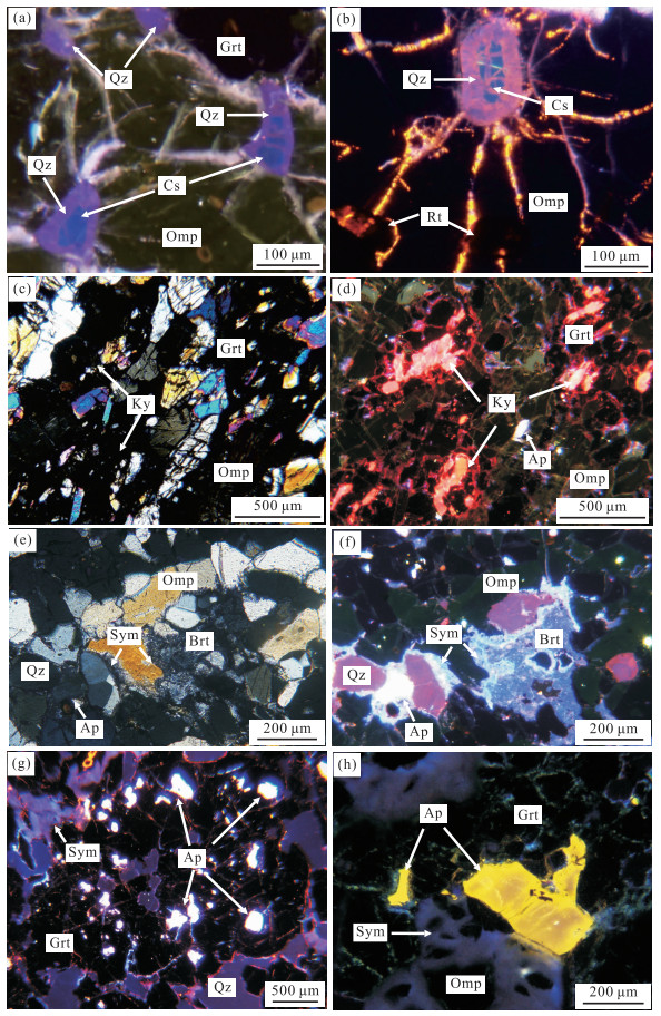
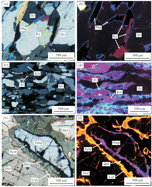
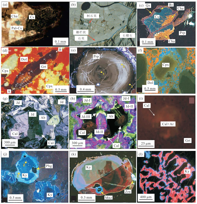
| Citation: | Wang Songjie, Wang Lu, Fu Jianmin, Ding Yue, 2014. A New Perspective for Research of Dabie-Sulu Ultrahigh-Pressure Metamorphic Rocks: Application of Optical Microscope-Based Cathodoluminescence. Earth Science, 39(3): 357-367. doi: 10.3799/dqkx.2014.034

|

|
Barker, C.E., Kopp, O.C., Colburn, H.Y., 1991. Luminescence Microscopy and Spectroscopy: Qualitative and Quantitative Applications: Text for Short Course No. 25 Sponsored by the Society of Economic Paleontologists and Mineralogists. SEPM, Dallas.
|
|
Dobrzhinetskaya, L.F., Wirth, R., GreenⅡ, H.W., 2006. Nanometric Inclusions of Carbonates in Kokchetav Diamonds from Kazakhstan: A New Constraint for the Depth of Metamorphic Diamond Crystallization. Earth and Planetary Science Letters, 243(1-2): 85-93. doi:org/ 10.1016/j.epsl.2005.11.030
|
|
Fu, Y.H., 2011. Cathodoluminescence Technology and Its Application in the Research of Sedimentary Rock's Cement. Petrochemical Industry Application, 30(8): 22-25 (in Chinese with English abstract). http://en.cnki.com.cn/Article_en/CJFDTOTAL-NXSH201108013.htm
|
|
Gaft, M., Reisfeld, R., Panczer, G., 2005. Modern Luminescence Spectroscopy of Minerals and Materials. Springer, Berlin.
|
|
Götze, J., 2002. Potential of Cathodoluminescence (CL) Microscopy and Spectroscopy for the Analysis of Minerals and Materials. Analytical and Bioanalytical Chemistry, 374(4): 703-708. doi: 10.1007/s00216-002-1461-1
|
|
Götze, J., 2012. Applications of Cathodoluminescence Microscopy and Spectroscopy in Geosciences. Microscopy and Microanalysis, 18(6): 1270-1284. doi: 10.1017/S1431927612001122
|
|
Götze, J., Kempe, U., 2008. A Comparison of Optical Microscope- and Scanning Electron Microscope-Based Cathodoluminescence (CL) Imaging and Spectroscopy Applied to Geosciences. Mineralogical Magazine, 72(4): 909-924. doi:org/ 10.1180/minmag.2008.072.4.909
|
|
Götze, J., Magnus, M., 1997. Quantitative Determination of Mineral Abundance in Geological Samples Using Combined Cathodoluminescence Microscopy and Image Analysis. European Journal of Mineralogy, 9(6): 1207-1215. doi: 10.1127/ejm/9/6/1207
|
|
Götze, J., Schertl, H.P., Neuser, R.D., et al., 2012. Optical Microscope-Cathodoluminescence (OM-CL) Imaging as a Powerful Tool to Reveal Internal Textures of Minerals. Mineralogy and Petrology, 107(3): 373-392. doi: 10.1007/s00710-012-0256-0
|
|
Gucsik, A., 2009. Cathodoluminescence and Its Application in the Planetary Sciences. Springer, Berlin.
|
|
Huang, S.J., 1992. Relationship between Cathodoluminescence and Concentration of Iron and Manganese in Carbonate Minerals. Mineralogy and Petrology, 12(4): 74-79 (in Chinese with English abstract). http://en.cnki.com.cn/Article_en/CJFDTOTAL-KWYS199204010.htm
|
|
Huang, S.J., Qing, H.R., Hu, Z.W., et al., 2008. Cathodoluminescence and Diagenesis of the Carbonate Rocks in Feixianguan Formation of Trassic, Eastern Sichuan Basin of China. Earth Science—Journal of China University of Geosciences, 33(1): 26-34 (in Chinese with English abstract). doi: 10.3799/dqkx.2008.004
|
|
Ingrin, J., Gillet, P., 1986. TEM Investigation of the Crystal Microstructures in a Quartz-Coesite Assemblage of the Western Alps. Physics and Chemistry of Minerals, 13(5): 325-330. doi: 10.1007/BF00308349
|
|
Korsakov, A.V., De Gussem, K., Zhukov, V.P., et al., 2009. Aragonite-Calcite-Dolomite Relationships in UHPM Polycrystalline Carbonate Inclusions from the Kokchetav Massif, Northern Kazakhstan. European Journal of Mineralogy, 21(6): 1301-1311. doi: 10.1127/0935-1221/2009/0021-1992
|
|
Lai, Y., 1995. Application of Cathodoluminescence to Mineralization and Lithogenesis Studying. Acta Scientiarum Naturalium Universitatis Pekinensis, 31(5): 631-638 (in Chinese with English abstract). http://en.cnki.com.cn/Article_en/CJFDTOTAL-BJDZ505.016.htm
|
|
Langenhorst, F., Poirier, J.P., 2002. Transmission Electron Microscopy of Coesite Inclusions in the Dora Maira High-Pressure Metamorphic Pyrope-Quartzite. Earth and Planetary Science Letters, 203(3-4): 793-803. doi:org/ 10.1016/S0012-821X(02)00949-4
|
|
Lenze, A., Stöckhert, B., 2008. Microfabrics of Quartz Formed from Coesite (Dora-Maira Massif, Western Alps). European Journal of Mineralogy, 20(5): 811-826. doi: 10.1127/0935-1221/2008/0020-1848
|
|
Li, S.G., Li, H.M., Chen, Y.Z., et al., 1997. The UHP Metamorphic Geochronology of Dabie-Sulu Terrain—Ⅱ. Zircon U-Pb Isotope System. Science in China (Series D), 27(3): 200-206 (in Chinese).
|
|
Liu, J., Huangfu, H.Y., 2000. The Cathodoluminescence and Trace Elements in Carbonate Minerals. Sedimentary Geology and Tethyan Geology, 20(3): 71-76 (in Chinese with English abstract). http://www.cnki.com.cn/Article/CJFDTotal-TTSD200003012.htm
|
|
Maresch, W.V., Grevel, C., Stanek, K.P., et al., 2012. Multiple Growth Mechanisms of Jadeite in Cuban Metabasite. European Journal of Mineralogy, 24(2): 217-235. doi: 10.1127/0935-1221/2012/0024-2179
|
|
Marfunin, A.S., 1979. Spectroscopy, Luminescence and Radiation Centers in Minerals. Springer, Berlin.
|
|
Marshall, D.J., Mariano, A.N., 1988. Cathodoluminescence of Geological Materials. Unwin Hyman, Boston.
|
|
Mosenfelder, J.L., Schertl, H.P., Smyth, J.R., et al., 2005. Factors in the Preservation of Coesite: The Importance of Fluid Infiltration. American Mineralogist, 90(5-6): 779-789. doi: 10.2138/am.2005.1687
|
|
Neuser, R.D., 1995. A New High-Intensity Cathodoluminescence Microscope and Its Application to Weakly Luminescing Minerals. Bochumer Geologische and Geotechnische Arbeiten, 44: 116-118. http://www.researchgate.net/profile/Rolf_Neuser/publication/280621081_A_new_high-intensity_cathodoluminescence_microscope_and_its_application_to_weakly_luminescing_minerals/links/55bf38f908aec0e5f445f50f.pdf
|
|
Pagel, M., Barbin, V., Blanc, P., et al., 2000. Cathodoluminescence in Geosciences. Springer, Berlin.
|
|
Peng, H.J., Wang, X.W., Tang, J.X., et al., 2010. The Application of Quartz Cathodoluminescence in Study of Igneous Rock. Rock and Mineral Analysis, 29(2): 153-160 (in Chinese with English abstract). http://en.cnki.com.cn/Article_en/CJFDTOTAL-YKCS201002018.htm
|
|
Ramseyer, K., AIDahan, A.A., Collini, B., et al., 1992. Petrological Modifications in Granitic Rocks from the Siljan Impact Structure: Evidence from Cathodoluminescence. Tectonophysics, 216(1-2): 195-204. doi:org/ 10.1016/0040-1951(92)90166-4
|
|
Richter, D.K., Götte, T., Götze, J., et al., 2003. Progress in Application of Cathodoluminescence (CL) in Sedimentary Petrology. Mineralogy and Petrology, 79(3-4): 127-166. doi: 10.1007/s00710-003-0237-4
|
|
Schertl, H.P., Maresch, W.V., Stanek, K.P., et al., 2012. New Occurrences of Jadeitite, Jadeite Quartzite and Jadeite-Lawsonite Quartzite in the Dominican Republic, Hispaniola: Petrological and Geochronological Overview. European Journal of Mineralogy, 24(2): 199-216. doi: 10.1127/0935-1221/2012/0024-2201
|
|
Schertl, H.P., Medenbach, O., Neuser, R.D., 2005. UHP-Metamorphic Rocks from Dora Maira, Western Alps: Cathodoluminescence of Silica and Twinning of Coesite. Russian Geology and Geophysics, 46(12): 1345-1351. http://www.researchgate.net/publication/234058050_UHP-metamorphic_rocks_from_Dora_Maira_Western_Alps_Cathodoluminescence_of_silica_and_twinning_of_coesite
|
|
Schertl, H.P., Neuser, R.D., Sobolev, N.V., et al., 2004. UHP-Metamorphic Rocks from Dora Maria/Western Alps and Kokchetav/Kazakhstan: New Insights Using Cathodoluminescence Petrology. European Journal of Mineralogy, 16(1): 49-57. doi: 10.1127/0935-1221/2004/0016-0049
|
|
Sippel, R.F., 1965. Simple Device for Luminescence Petrology. Review of Scientific Instruments, 36(11): 556-558.
|
|
Sippel, R.F., Glover, E.D., 1965. Structures in Carbonate Rocks Made Visible by Luminescence Petrology. Science, 150(3701): 1283-1287. doi: 10.1126/science.150.3701.1283
|
|
Sobolev, N.V., Schertl, H.P., Neuser, R.D., et al., 2007. Relict Unusually Low Iron Pyrope-Grossular Garnets in UHPM Calc-Silicate Rocks of the Kokchetav Massif, Kazakhstan. International Geology Review, 49(8): 717-731. doi: 10.2747/0020-6814.49.8.717
|
|
Sobolev, N.V., Schertl, H.P., Valley, J.W., et al., 2011. Oxygen Isotope Variations of Garnets and Clinopyroxenes in a Layered Diamondiferous Calcsilicate Rock from Kokchetav Massif, Kazakhstan: A Window into the Geochemical Nature of Deeply Subducted UHPM Rocks. Contributions to Mineralogy and Petrology, 162(5): 1079-1092. doi: 10.1007/s00410-011-0641-4
|
|
Sun, J., Huang, X.P., Jin, Z.K., et al., 2009. Controlling Factors of Cathodoluminescence of Carbonate Minerals. Sedimentary Geology and Tethyan Geology, 29(1): 102-108 (in Chinese with English abstract).
|
|
Tian, H.J., 1989. The Application of Cathodoluminescence in Sedimentology. Sedimentary Facies and Palaeogeography, 43(5): 56-65 (in Chinese).
|
|
Wang, L., Jin, Z.M., Kusky, T.M., et al., 2010a. Microfabric Characteristics and Rheological Significance of Ultra-High-Pressure Metamorphosed Jadeite-Quartzite and Eclogite from Shuanghe, Dabie Mountains, China. Journal of Metamorphic Geology, 28(2): 163-182. doi: 10.1111/j.1525-1314.2009.00859.x
|
|
Wang, L., Kusky, T.M., Li, S.Z., 2010b. Structural Geometry of an Exhumed UHP Terrane in the Eastern Sulu Orogen, China: Implications for Continental Collisional Processes. Journal of Structural Geology, 32(4): 423-444. doi:org/ 10.1016/j.jsg.2010.01.012
|
|
Wang, Y.Q., Zhang, S.P., Ying, F.X., 1996. Applications of Cathodoluminescence Microscopy in Reservoir Research. Petroleum Industry Press, Beijing (in Chinese).
|
|
Wu, Y.B., Zheng, Y.F., 2004. Genesis of Zircon and Its Constraints on Interpretation of U-Pb Age. Chinese Science Bulletin, 49(16): 1589-1604 (in Chinese). doi: 10.1360/csb2004-49-16-1589
|
|
Xu, H.F., Chen, T., 1987. Application of Cathodoluminescence to Metamorphic and Granitic Rocks. Acta Petrologica et Mineralogica, 6(3): 279-284 (in Chinese with English abstract). http://en.cnki.com.cn/Article_en/CJFDTOTAL-YSKW198703008.htm
|
|
Xu, H.F., Cui, J.G., Qiu, X.P., 2006. Applications of Cathodoluminescence Technology in Petrology and Mineral Deposits. Geological Publishing House, Beijing (in Chinese).
|
|
Yang, Y., Chen, N.S., 2003. UV-Cathodoluminescence Mechanism of Secondary Enlarged Quartz and Its Importance. Rock and Mineral Analysis, 22(1): 1-3 (in Chinese with English abstract). http://en.cnki.com.cn/Article_en/CJFDTOTAL-YKCS200301001.htm
|
|
Ying, F.X., Wang, Y.Q., 1990. Elemental Composition and CL Color of Minerals. Journal of Chinese Electron Microscopy Society, 9(3): 244-244 (in Chinese).
|
|
Yu, B.S., 1992. The Application and Development of Cathodoluminescence Microscope in the Study of Carbonate Rocks. Geological Science and Technology Information, 11(4): 92-96 (in Chinese with English abstract). http://en.cnki.com.cn/Article_en/CJFDTOTAL-DZKQ199204020.htm
|
|
Zhang, B.Q., Yu, H.Z., Jiang, Z.X., et al., 2003. Characteristics and Diagenetic Environments of Source Rocks by Cathodoluminescence. Petroleum Exploration and Development, 6: 117-120 (in Chinese with English abstract). http://en.cnki.com.cn/Article_en/CJFDTOTAL-SKYK200303033.htm
|
|
Zheng, Y.F., Zhao, Z.F., Wu, Y.B., et al., 2006. Zircon U-Pb Age, Hf and O Isotope Constraints on Protolith Origin of Ultrahigh-Pressure Eclogite and Gneiss in the Dabie Orogen. Chemical Geology, 231(1-2): 135-158. doi:org/ 10.1016/j.chemgeo.2006.01.005
|
|
付月红, 2011. 阴极发光技术在研究沉积岩胶结物中的应用. 石油化工应用, 30(8): 22-25. doi: 10.3969/j.issn.1673-5285.2011.08.008
|
|
黄思静, 1992. 碳酸盐矿物的阴极发光性与其Fe、Mn含量的关系. 矿物岩石, 12(4): 74-79. https://www.cnki.com.cn/Article/CJFDTOTAL-KWYS199204010.htm
|
|
黄思静, 卿海若, 胡作维, 等, 2008. 川东三叠系飞仙关组碳酸盐岩的阴极发光特征与成岩作用. 地球科学——中国地质大学学报, 33(1): 26-34. https://www.cnki.com.cn/Article/CJFDTOTAL-DQKX200801007.htm
|
|
赖勇, 1995. 阴极发光技术在成岩成矿作用研究中的应用. 北京大学学报(自然科学版), 5: 631-638. https://www.cnki.com.cn/Article/CJFDTOTAL-BJDZ505.016.htm
|
|
李曙光, 李惠民, 陈移之, 等, 1997. 大别山-苏鲁地体超高压变质年代学——Ⅱ. 锆石U-Pb同位素体系. 中国科学(D辑), 27(3): 200-206. https://www.cnki.com.cn/Article/CJFDTOTAL-JDXK199703001.htm
|
|
刘洁, 皇甫红英, 2000. 碳酸盐矿物的阴极发光性与微量元素的关系. 沉积与特提斯地质, 20(3): 71-76. doi: 10.3969/j.issn.1009-3850.2000.03.013
|
|
彭惠娟, 汪雄武, 唐菊兴, 等, 2010. 石英阴极发光在火成岩研究中的应用. 岩矿测试, 29(2): 153-160. doi: 10.3969/j.issn.0254-5357.2010.02.014
|
|
孙靖, 黄小平, 金振奎, 等, 2009. 碳酸盐矿物阴极发光性的控制因素分析. 沉积与特提斯地质, 29(1): 102-108. doi: 10.3969/j.issn.1009-3850.2009.01.017
|
|
田洪均, 1989. 阴极发光技术在沉积学中的应用. 岩相古地理, 43(5): 56-65. https://www.cnki.com.cn/Article/CJFDTOTAL-TTSD198905006.htm
|
|
王衍琦, 张绍平, 应凤祥, 1996. 阴极发光显微镜在储层研究中的应用. 北京: 石油工业出版社.
|
|
吴元保, 郑永飞, 2004. 锆石成因矿物学研究及其对U-Pb年龄解释的制约. 科学通报, 49(16): 1589-1604. doi: 10.3321/j.issn:0023-074X.2004.16.002
|
|
徐惠芬, 陈涛, 1987. 阴极发光仪在变质岩和花岗岩类岩石中的应用. 岩石矿物学杂志, 6(3): 279-284. https://www.cnki.com.cn/Article/CJFDTOTAL-YSKW198703008.htm
|
|
徐惠芬, 崔京钢, 邱小平, 2006. 阴极发光技术在岩石学和矿床学中的应用. 北京: 地质出版社.
|
|
杨勇, 陈能松, 2003. 次生石英的紫外阴极发光机理及意义. 岩矿测试, 22(1): 1-3. doi: 10.3969/j.issn.0254-5357.2003.01.001
|
|
应凤祥, 王衍琦, 1990. 矿物的元素组成与阴极发光颜色. 电子显微学报, 9(3): 244-244. https://www.cnki.com.cn/Article/CJFDTOTAL-DZXV199003243.htm
|
|
于炳松, 1992. 阴极发光显微镜在碳酸盐岩研究中的应用及进展. 地质科技情报, 11(4): 92-96. https://www.cnki.com.cn/Article/CJFDTOTAL-DZKQ199204020.htm
|
|
张本琪, 余宏忠, 姜在兴, 等, 2003. 应用阴极发光技术研究母岩性质及成岩环境. 石油勘探与开发, 6: 117-120. https://www.cnki.com.cn/Article/CJFDTOTAL-SKYK200303033.htm
|
Figures(4)

 DownLoad: