


A New Perspective for Research of Dabie-Sulu Ultrahigh-Pressure Metamorphic Rocks: Application of Optical Microscope-Based Cathodoluminescence
-
摘要: 利用偏光显微镜阴极发光技术可观察到其他常规成分结构测试法不易识别或容易忽略的多种矿物的生长结构, 该技术是进行后续成分分析的有效预研究手段, 可为重建矿物形成演化过程提供重要信息.该技术在国际岩石矿物学、油气储层及矿床学领域应用广泛, 但在变质岩研究领域的应用较薄弱.综述该技术在国际超高压变质岩研究领域的应用, 并利用其对大别-苏鲁超高压变质带经典地区的超高压榴辉岩、云母片岩、大理岩进行初步研究, 讨论它在多期微细矿物相快速鉴别、生长环带、微量元素分布规律、双晶纹、出溶结构等内部结构表征方面的应用价值和前景.偏光显微镜阴极发光技术与拉曼光谱、扫描电镜、电子探针等分析技术相结合, 可为我国超高压变质岩的研究开辟和扩展一条新思路.Abstract: With the use of optical microscope-based cathodoluminescence (OM-CL), many kinds of growth textures of minerals can be observed, which are either indiscernable or to be omitted easily with other routine analytical methods. OM-CL is an effective pre-research technique prior to other follow-up component analysis, which can provide important information for reconstructing formation and evolution processes of minerals. This technique has wide-spread applications in international petromineralogy, oil and gas reservoir and mineral deposits, but is relatively weak in metamorphic rocks. The applications of OM-CL in the UHP (ultra high power) metamorphic rocks are reviewed in this paper, as well as preliminary studies by OM-CL on ultrahigh-pressure eclogites, micaschist and marble at classical areas in the Dabie-Sulu UHP metamorphic belt. And its application and prospect in the fast identification of multi-phase tiny mineral facies and internal structural characterization, including growth zoning, distribution of trace elements, twinning and exsolution texture are discussed. A new perspective for our research on ultrahigh-pressure metamorphic rocks can be developed when combining OM-CL with mineral chemistry analysis techniques such as Roman spectrum, scanning electron microscope (SEM) and electron probe micro-analyzer (EPMA).
-
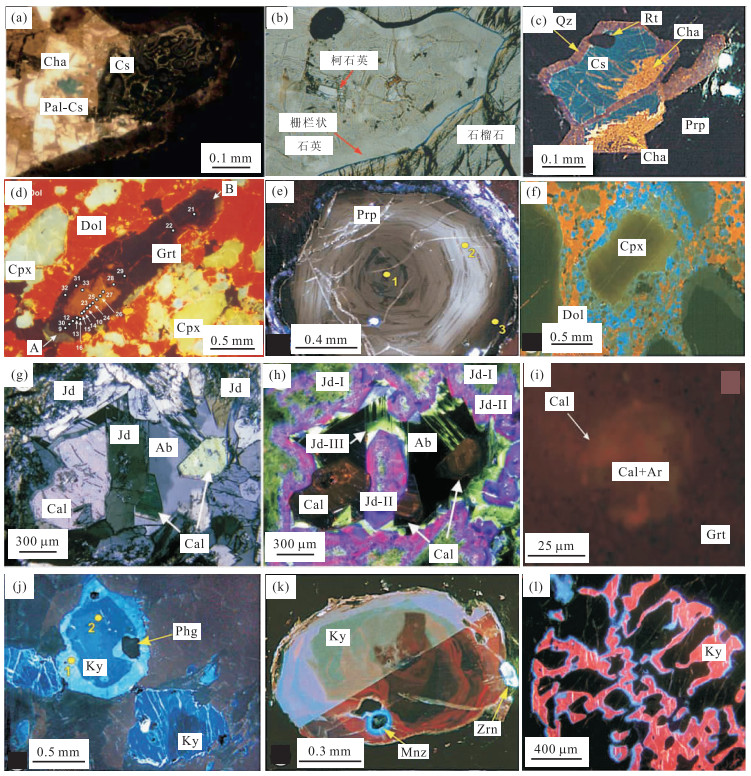
图 1 各超高压-高压变质带中不同矿物的典型阴极发光图像(引自Schertl et al., 2004, 2012; Mosenfelder et al., 2005; Sobolev et al., 2011; Götze et al., 2012)
a~c为柯石英及假象的阴极发光(a, c)和单偏光(b)图像,CL下柯石英(蓝色)-栅状石英(紫罗兰色)-玉髓(黄棕色);d~e为镁铝榴石阴极发光图像,d显示核部和边部发光强度不同,e显示出典型生长环带;f为透辉石阴极发光图像,核部呈绿色,边部为蓝色;g~h为硬玉显微特征对比,CL可以显示出3期硬玉生长阶段;i为方解石+霰石多相包裹体阴极发光图像;j, k, l分别为蓝晶石生长环带、双晶纹和海绵状生长结构.其中,Ab.钠长石;Ar.文石;Cal.方解石;Cha.玉髓;Cpx.单斜辉石;Cs.柯石英;Dol.白云石;Grt.石榴石;Jd.硬玉;Ky.蓝晶石;Mnz.独居石;Pal-Cs.栅状石英;Phg.多硅白云母;Prp.镁铝榴石;Qz.石英;Rt.金红石;Zrn.锆石
Fig. 1. Typical CL microphotographs of different minerals from several UHP-HP metamorphic belts
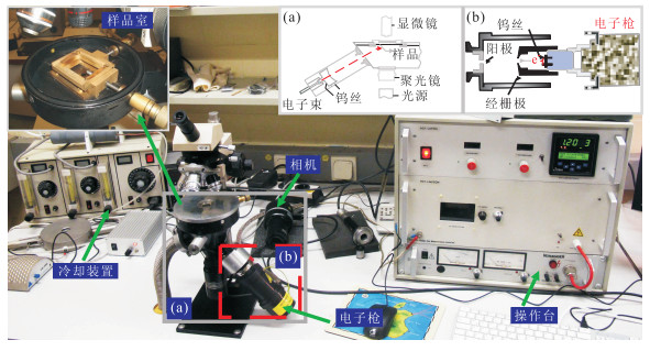
图 2 德国波鸿大学HC1-CM型“热阴极”偏光显微镜发光仪主要部件组成(部分修编于Götze and Kempe, 2008)
a, b分别为“热阴极”发光仪内部配置和电子枪构成
Fig. 2. Principle components of type HC1-CM hot-cathode microscope developed at the Ruhr-University, Bochum
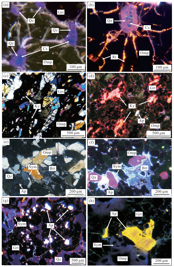
图 3 大别-苏鲁地区不同类型榴辉岩偏光显微照片与阴极发光特征对比
a, b.仰口24b和双河SH2样品中柯石英及其假象的阴极发光图像,柯石英呈蓝色,栅状石英为紫罗兰色;c, d.YK3样品中蓝晶石的正交光和CL图像,蓝晶石在偏光镜下(c)特征不明显,而在CL下(d)则为鲜红色,分布特征可以清楚识别;e, f.重晶石的显微特征对比,正交镜下(e)特征不明显,而CL下(f)表现为蓝色;g.SH2中磷灰石包裹体CL图像,磷灰石表现为亮白色;h.退变质榴辉岩(YK15)中磷灰石CL图像,磷灰石呈黄色,边部和核部发光强度不同,核部颜色更暗.其中,Omp.绿辉石;Brt.重晶石;Ap.磷灰石;Sym.后成合晶;其他见图 1
Fig. 3. Comparison of cathodoluminescence characteristics with polarized micrographs for different types of eclogites in Dabie-Sulu area
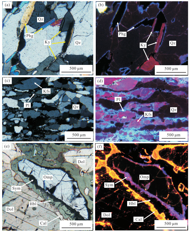
图 4 云母片岩、长英质片麻岩和绿辉石大理岩显微特征对比
a, b.含蓝晶石云母片岩正交偏光图像(a)与阴极发光图像(b)对比,CL下蓝晶石为鲜红色,石英呈暗棕红色,云母不发光;c, d.长英质片麻岩正交偏光图像(c)与阴极发光图像(d)对比,石英发暗棕红色光,长石呈蓝-紫色和浅蓝色;e, f.绿辉石大理岩典型显微特征对比,CL下(f)可看到角闪石和白云石之间的鲜黄色反应边.其中,Kfs.钾长石;Pl.斜长石;Hbl.角闪石;其他见图 1及图 3
Fig. 4. Comparison of microscopic features for micaschist, quartz-feldspathic gneiss and marble
-
Barker, C.E., Kopp, O.C., Colburn, H.Y., 1991. Luminescence Microscopy and Spectroscopy: Qualitative and Quantitative Applications: Text for Short Course No. 25 Sponsored by the Society of Economic Paleontologists and Mineralogists. SEPM, Dallas. Dobrzhinetskaya, L.F., Wirth, R., GreenⅡ, H.W., 2006. Nanometric Inclusions of Carbonates in Kokchetav Diamonds from Kazakhstan: A New Constraint for the Depth of Metamorphic Diamond Crystallization. Earth and Planetary Science Letters, 243(1-2): 85-93. doi:org/ 10.1016/j.epsl.2005.11.030 Fu, Y.H., 2011. Cathodoluminescence Technology and Its Application in the Research of Sedimentary Rock's Cement. Petrochemical Industry Application, 30(8): 22-25 (in Chinese with English abstract). http://en.cnki.com.cn/Article_en/CJFDTOTAL-NXSH201108013.htm Gaft, M., Reisfeld, R., Panczer, G., 2005. Modern Luminescence Spectroscopy of Minerals and Materials. Springer, Berlin. Götze, J., 2002. Potential of Cathodoluminescence (CL) Microscopy and Spectroscopy for the Analysis of Minerals and Materials. Analytical and Bioanalytical Chemistry, 374(4): 703-708. doi: 10.1007/s00216-002-1461-1 Götze, J., 2012. Applications of Cathodoluminescence Microscopy and Spectroscopy in Geosciences. Microscopy and Microanalysis, 18(6): 1270-1284. doi: 10.1017/S1431927612001122 Götze, J., Kempe, U., 2008. A Comparison of Optical Microscope- and Scanning Electron Microscope-Based Cathodoluminescence (CL) Imaging and Spectroscopy Applied to Geosciences. Mineralogical Magazine, 72(4): 909-924. doi:org/ 10.1180/minmag.2008.072.4.909 Götze, J., Magnus, M., 1997. Quantitative Determination of Mineral Abundance in Geological Samples Using Combined Cathodoluminescence Microscopy and Image Analysis. European Journal of Mineralogy, 9(6): 1207-1215. doi: 10.1127/ejm/9/6/1207 Götze, J., Schertl, H.P., Neuser, R.D., et al., 2012. Optical Microscope-Cathodoluminescence (OM-CL) Imaging as a Powerful Tool to Reveal Internal Textures of Minerals. Mineralogy and Petrology, 107(3): 373-392. doi: 10.1007/s00710-012-0256-0 Gucsik, A., 2009. Cathodoluminescence and Its Application in the Planetary Sciences. Springer, Berlin. Huang, S.J., 1992. Relationship between Cathodoluminescence and Concentration of Iron and Manganese in Carbonate Minerals. Mineralogy and Petrology, 12(4): 74-79 (in Chinese with English abstract). http://en.cnki.com.cn/Article_en/CJFDTOTAL-KWYS199204010.htm Huang, S.J., Qing, H.R., Hu, Z.W., et al., 2008. Cathodoluminescence and Diagenesis of the Carbonate Rocks in Feixianguan Formation of Trassic, Eastern Sichuan Basin of China. Earth Science—Journal of China University of Geosciences, 33(1): 26-34 (in Chinese with English abstract). doi: 10.3799/dqkx.2008.004 Ingrin, J., Gillet, P., 1986. TEM Investigation of the Crystal Microstructures in a Quartz-Coesite Assemblage of the Western Alps. Physics and Chemistry of Minerals, 13(5): 325-330. doi: 10.1007/BF00308349 Korsakov, A.V., De Gussem, K., Zhukov, V.P., et al., 2009. Aragonite-Calcite-Dolomite Relationships in UHPM Polycrystalline Carbonate Inclusions from the Kokchetav Massif, Northern Kazakhstan. European Journal of Mineralogy, 21(6): 1301-1311. doi: 10.1127/0935-1221/2009/0021-1992 Lai, Y., 1995. Application of Cathodoluminescence to Mineralization and Lithogenesis Studying. Acta Scientiarum Naturalium Universitatis Pekinensis, 31(5): 631-638 (in Chinese with English abstract). http://en.cnki.com.cn/Article_en/CJFDTOTAL-BJDZ505.016.htm Langenhorst, F., Poirier, J.P., 2002. Transmission Electron Microscopy of Coesite Inclusions in the Dora Maira High-Pressure Metamorphic Pyrope-Quartzite. Earth and Planetary Science Letters, 203(3-4): 793-803. doi:org/ 10.1016/S0012-821X(02)00949-4 Lenze, A., Stöckhert, B., 2008. Microfabrics of Quartz Formed from Coesite (Dora-Maira Massif, Western Alps). European Journal of Mineralogy, 20(5): 811-826. doi: 10.1127/0935-1221/2008/0020-1848 Li, S.G., Li, H.M., Chen, Y.Z., et al., 1997. The UHP Metamorphic Geochronology of Dabie-Sulu Terrain—Ⅱ. Zircon U-Pb Isotope System. Science in China (Series D), 27(3): 200-206 (in Chinese). Liu, J., Huangfu, H.Y., 2000. The Cathodoluminescence and Trace Elements in Carbonate Minerals. Sedimentary Geology and Tethyan Geology, 20(3): 71-76 (in Chinese with English abstract). http://www.cnki.com.cn/Article/CJFDTotal-TTSD200003012.htm Maresch, W.V., Grevel, C., Stanek, K.P., et al., 2012. Multiple Growth Mechanisms of Jadeite in Cuban Metabasite. European Journal of Mineralogy, 24(2): 217-235. doi: 10.1127/0935-1221/2012/0024-2179 Marfunin, A.S., 1979. Spectroscopy, Luminescence and Radiation Centers in Minerals. Springer, Berlin. Marshall, D.J., Mariano, A.N., 1988. Cathodoluminescence of Geological Materials. Unwin Hyman, Boston. Mosenfelder, J.L., Schertl, H.P., Smyth, J.R., et al., 2005. Factors in the Preservation of Coesite: The Importance of Fluid Infiltration. American Mineralogist, 90(5-6): 779-789. doi: 10.2138/am.2005.1687 Neuser, R.D., 1995. A New High-Intensity Cathodoluminescence Microscope and Its Application to Weakly Luminescing Minerals. Bochumer Geologische and Geotechnische Arbeiten, 44: 116-118. http://www.researchgate.net/profile/Rolf_Neuser/publication/280621081_A_new_high-intensity_cathodoluminescence_microscope_and_its_application_to_weakly_luminescing_minerals/links/55bf38f908aec0e5f445f50f.pdf Pagel, M., Barbin, V., Blanc, P., et al., 2000. Cathodoluminescence in Geosciences. Springer, Berlin. Peng, H.J., Wang, X.W., Tang, J.X., et al., 2010. The Application of Quartz Cathodoluminescence in Study of Igneous Rock. Rock and Mineral Analysis, 29(2): 153-160 (in Chinese with English abstract). http://en.cnki.com.cn/Article_en/CJFDTOTAL-YKCS201002018.htm Ramseyer, K., AIDahan, A.A., Collini, B., et al., 1992. Petrological Modifications in Granitic Rocks from the Siljan Impact Structure: Evidence from Cathodoluminescence. Tectonophysics, 216(1-2): 195-204. doi:org/ 10.1016/0040-1951(92)90166-4 Richter, D.K., Götte, T., Götze, J., et al., 2003. Progress in Application of Cathodoluminescence (CL) in Sedimentary Petrology. Mineralogy and Petrology, 79(3-4): 127-166. doi: 10.1007/s00710-003-0237-4 Schertl, H.P., Maresch, W.V., Stanek, K.P., et al., 2012. New Occurrences of Jadeitite, Jadeite Quartzite and Jadeite-Lawsonite Quartzite in the Dominican Republic, Hispaniola: Petrological and Geochronological Overview. European Journal of Mineralogy, 24(2): 199-216. doi: 10.1127/0935-1221/2012/0024-2201 Schertl, H.P., Medenbach, O., Neuser, R.D., 2005. UHP-Metamorphic Rocks from Dora Maira, Western Alps: Cathodoluminescence of Silica and Twinning of Coesite. Russian Geology and Geophysics, 46(12): 1345-1351. http://www.researchgate.net/publication/234058050_UHP-metamorphic_rocks_from_Dora_Maira_Western_Alps_Cathodoluminescence_of_silica_and_twinning_of_coesite Schertl, H.P., Neuser, R.D., Sobolev, N.V., et al., 2004. UHP-Metamorphic Rocks from Dora Maria/Western Alps and Kokchetav/Kazakhstan: New Insights Using Cathodoluminescence Petrology. European Journal of Mineralogy, 16(1): 49-57. doi: 10.1127/0935-1221/2004/0016-0049 Sippel, R.F., 1965. Simple Device for Luminescence Petrology. Review of Scientific Instruments, 36(11): 556-558. Sippel, R.F., Glover, E.D., 1965. Structures in Carbonate Rocks Made Visible by Luminescence Petrology. Science, 150(3701): 1283-1287. doi: 10.1126/science.150.3701.1283 Sobolev, N.V., Schertl, H.P., Neuser, R.D., et al., 2007. Relict Unusually Low Iron Pyrope-Grossular Garnets in UHPM Calc-Silicate Rocks of the Kokchetav Massif, Kazakhstan. International Geology Review, 49(8): 717-731. doi: 10.2747/0020-6814.49.8.717 Sobolev, N.V., Schertl, H.P., Valley, J.W., et al., 2011. Oxygen Isotope Variations of Garnets and Clinopyroxenes in a Layered Diamondiferous Calcsilicate Rock from Kokchetav Massif, Kazakhstan: A Window into the Geochemical Nature of Deeply Subducted UHPM Rocks. Contributions to Mineralogy and Petrology, 162(5): 1079-1092. doi: 10.1007/s00410-011-0641-4 Sun, J., Huang, X.P., Jin, Z.K., et al., 2009. Controlling Factors of Cathodoluminescence of Carbonate Minerals. Sedimentary Geology and Tethyan Geology, 29(1): 102-108 (in Chinese with English abstract). Tian, H.J., 1989. The Application of Cathodoluminescence in Sedimentology. Sedimentary Facies and Palaeogeography, 43(5): 56-65 (in Chinese). Wang, L., Jin, Z.M., Kusky, T.M., et al., 2010a. Microfabric Characteristics and Rheological Significance of Ultra-High-Pressure Metamorphosed Jadeite-Quartzite and Eclogite from Shuanghe, Dabie Mountains, China. Journal of Metamorphic Geology, 28(2): 163-182. doi: 10.1111/j.1525-1314.2009.00859.x Wang, L., Kusky, T.M., Li, S.Z., 2010b. Structural Geometry of an Exhumed UHP Terrane in the Eastern Sulu Orogen, China: Implications for Continental Collisional Processes. Journal of Structural Geology, 32(4): 423-444. doi:org/ 10.1016/j.jsg.2010.01.012 Wang, Y.Q., Zhang, S.P., Ying, F.X., 1996. Applications of Cathodoluminescence Microscopy in Reservoir Research. Petroleum Industry Press, Beijing (in Chinese). Wu, Y.B., Zheng, Y.F., 2004. Genesis of Zircon and Its Constraints on Interpretation of U-Pb Age. Chinese Science Bulletin, 49(16): 1589-1604 (in Chinese). doi: 10.1360/csb2004-49-16-1589 Xu, H.F., Chen, T., 1987. Application of Cathodoluminescence to Metamorphic and Granitic Rocks. Acta Petrologica et Mineralogica, 6(3): 279-284 (in Chinese with English abstract). http://en.cnki.com.cn/Article_en/CJFDTOTAL-YSKW198703008.htm Xu, H.F., Cui, J.G., Qiu, X.P., 2006. Applications of Cathodoluminescence Technology in Petrology and Mineral Deposits. Geological Publishing House, Beijing (in Chinese). Yang, Y., Chen, N.S., 2003. UV-Cathodoluminescence Mechanism of Secondary Enlarged Quartz and Its Importance. Rock and Mineral Analysis, 22(1): 1-3 (in Chinese with English abstract). http://en.cnki.com.cn/Article_en/CJFDTOTAL-YKCS200301001.htm Ying, F.X., Wang, Y.Q., 1990. Elemental Composition and CL Color of Minerals. Journal of Chinese Electron Microscopy Society, 9(3): 244-244 (in Chinese). Yu, B.S., 1992. The Application and Development of Cathodoluminescence Microscope in the Study of Carbonate Rocks. Geological Science and Technology Information, 11(4): 92-96 (in Chinese with English abstract). http://en.cnki.com.cn/Article_en/CJFDTOTAL-DZKQ199204020.htm Zhang, B.Q., Yu, H.Z., Jiang, Z.X., et al., 2003. Characteristics and Diagenetic Environments of Source Rocks by Cathodoluminescence. Petroleum Exploration and Development, 6: 117-120 (in Chinese with English abstract). http://en.cnki.com.cn/Article_en/CJFDTOTAL-SKYK200303033.htm Zheng, Y.F., Zhao, Z.F., Wu, Y.B., et al., 2006. Zircon U-Pb Age, Hf and O Isotope Constraints on Protolith Origin of Ultrahigh-Pressure Eclogite and Gneiss in the Dabie Orogen. Chemical Geology, 231(1-2): 135-158. doi:org/ 10.1016/j.chemgeo.2006.01.005 付月红, 2011. 阴极发光技术在研究沉积岩胶结物中的应用. 石油化工应用, 30(8): 22-25. doi: 10.3969/j.issn.1673-5285.2011.08.008 黄思静, 1992. 碳酸盐矿物的阴极发光性与其Fe、Mn含量的关系. 矿物岩石, 12(4): 74-79. https://www.cnki.com.cn/Article/CJFDTOTAL-KWYS199204010.htm 黄思静, 卿海若, 胡作维, 等, 2008. 川东三叠系飞仙关组碳酸盐岩的阴极发光特征与成岩作用. 地球科学——中国地质大学学报, 33(1): 26-34. https://www.cnki.com.cn/Article/CJFDTOTAL-DQKX200801007.htm 赖勇, 1995. 阴极发光技术在成岩成矿作用研究中的应用. 北京大学学报(自然科学版), 5: 631-638. https://www.cnki.com.cn/Article/CJFDTOTAL-BJDZ505.016.htm 李曙光, 李惠民, 陈移之, 等, 1997. 大别山-苏鲁地体超高压变质年代学——Ⅱ. 锆石U-Pb同位素体系. 中国科学(D辑), 27(3): 200-206. https://www.cnki.com.cn/Article/CJFDTOTAL-JDXK199703001.htm 刘洁, 皇甫红英, 2000. 碳酸盐矿物的阴极发光性与微量元素的关系. 沉积与特提斯地质, 20(3): 71-76. doi: 10.3969/j.issn.1009-3850.2000.03.013 彭惠娟, 汪雄武, 唐菊兴, 等, 2010. 石英阴极发光在火成岩研究中的应用. 岩矿测试, 29(2): 153-160. doi: 10.3969/j.issn.0254-5357.2010.02.014 孙靖, 黄小平, 金振奎, 等, 2009. 碳酸盐矿物阴极发光性的控制因素分析. 沉积与特提斯地质, 29(1): 102-108. doi: 10.3969/j.issn.1009-3850.2009.01.017 田洪均, 1989. 阴极发光技术在沉积学中的应用. 岩相古地理, 43(5): 56-65. https://www.cnki.com.cn/Article/CJFDTOTAL-TTSD198905006.htm 王衍琦, 张绍平, 应凤祥, 1996. 阴极发光显微镜在储层研究中的应用. 北京: 石油工业出版社. 吴元保, 郑永飞, 2004. 锆石成因矿物学研究及其对U-Pb年龄解释的制约. 科学通报, 49(16): 1589-1604. doi: 10.3321/j.issn:0023-074X.2004.16.002 徐惠芬, 陈涛, 1987. 阴极发光仪在变质岩和花岗岩类岩石中的应用. 岩石矿物学杂志, 6(3): 279-284. https://www.cnki.com.cn/Article/CJFDTOTAL-YSKW198703008.htm 徐惠芬, 崔京钢, 邱小平, 2006. 阴极发光技术在岩石学和矿床学中的应用. 北京: 地质出版社. 杨勇, 陈能松, 2003. 次生石英的紫外阴极发光机理及意义. 岩矿测试, 22(1): 1-3. doi: 10.3969/j.issn.0254-5357.2003.01.001 应凤祥, 王衍琦, 1990. 矿物的元素组成与阴极发光颜色. 电子显微学报, 9(3): 244-244. https://www.cnki.com.cn/Article/CJFDTOTAL-DZXV199003243.htm 于炳松, 1992. 阴极发光显微镜在碳酸盐岩研究中的应用及进展. 地质科技情报, 11(4): 92-96. https://www.cnki.com.cn/Article/CJFDTOTAL-DZKQ199204020.htm 张本琪, 余宏忠, 姜在兴, 等, 2003. 应用阴极发光技术研究母岩性质及成岩环境. 石油勘探与开发, 6: 117-120. https://www.cnki.com.cn/Article/CJFDTOTAL-SKYK200303033.htm -

 下载:
下载:
 点击查看大图
计量
- 文章访问数: 3501
- HTML全文浏览量: 1298
- PDF下载量: 355
- 被引次数: 0
