


NanoSIMS Techniques and Its Important Implications in Geomicrobiology and Biosedimentology
-
摘要: 纳米离子探针(NanoSIMS)技术具有高空间分辨率和高化学灵敏度的特点,是目前国际上最先进的原位微区分析手段之一.它能在纳米尺度上进行元素和同位素分布的原位分析,为揭示微生物与环境的相互作用提供了革命性技术;因此在地质微生物学和生物沉积学研究中展现出重要应用潜力甚至在某些方向的研究中不可或缺.本文在介绍NanoSIMS的工作原理基础上,综述了NanoSIMS技术在地质微生物学领域最前沿的重要应用,重点解剖其在现代微生物群落功能研究、深时碳酸盐矿物沉淀、生物地球化学循环、地球早期生命信号的识别以及地外生命探索等方面的应用实例.总之,NanoSIMS技术的发展与应用推动了地质微生物学研究迈向更高空间分辨率的微观世界,为探索地球生命起源和地外星球宜居性提供了前所未有的机遇.同时,本文论述了NanoSIMS在样品制备、信噪比、定量分析等方面的挑战,并展望了该技术优化方向,如技术升级、多技术联用和算法改进等,希望为多学科原位微区分析提供重要技术支撑,并进一步推动地质微生物学和生物沉积学及相关交叉学科的发展.Abstract: NanoSIMS (nanoscale secondary ion mass spectrometry) technology, with its high spatial resolution and chemical sensitivity, demonstrates significant potential for implications in researches of geomicrobiology. This technology enables in-situ analysis of elemental and isotopic distributions at the nanoscale, offering a revolutionary tool for uncovering interactions between microorganisms and their environments. This article reviews the applications and advancements of NanoSIMS technology in the field of geomicrobiology. It provides a detailed overview of the principles of NanoSIMS and its applications in areas such as identifying biogenic minerals from ancient microorganisms, exploring extraterrestrial life, carbonate mineral precipitation, biogeochemical cycles, and functional studies of modern microbial communities. The development of NanoSIMS has propelled geomicrobiology towards greater precision and resolution, presenting unprecedented opportunities to study the origin of life, microbial activity in extreme environments, and global biogeochemical cycles. At the same time, this article identifies challenges related to sample preparation, signal-to-noise ratio, and quantitative analysis, while also suggesting directions for optimization, such as technological upgrades, multi-technique integration, and algorithmic improvements. In the future, the continued evolution of NanoSIMS will provide critical support for investigating early life on Earth, global elemental cycles, and extraterrestrial life, further advancing geomicrobiology and its related interdisciplinary fields.
-
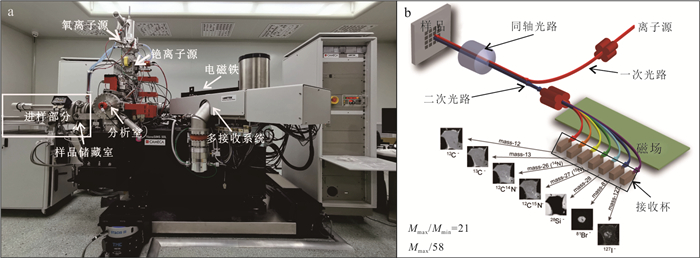
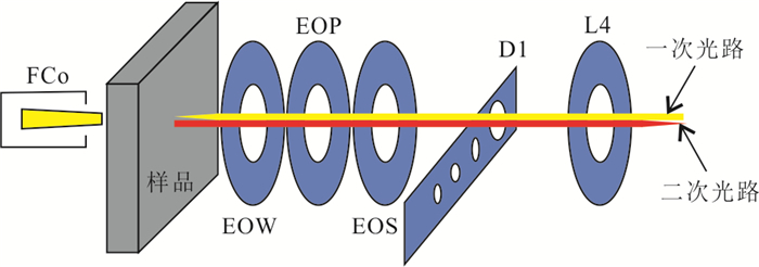
图 1 CAMECA NanoSIMS 50L型纳米离子探针设备(a)及其结构示意简图(b)
设备来自中国地质大学(武汉)地质微生物与环境全国重点实验室
Fig. 1. NanoSIMS (CAMECA NanoSIMS 50L) instrument (a) and the schematic diagram of the NanoSIMS 50L (b)
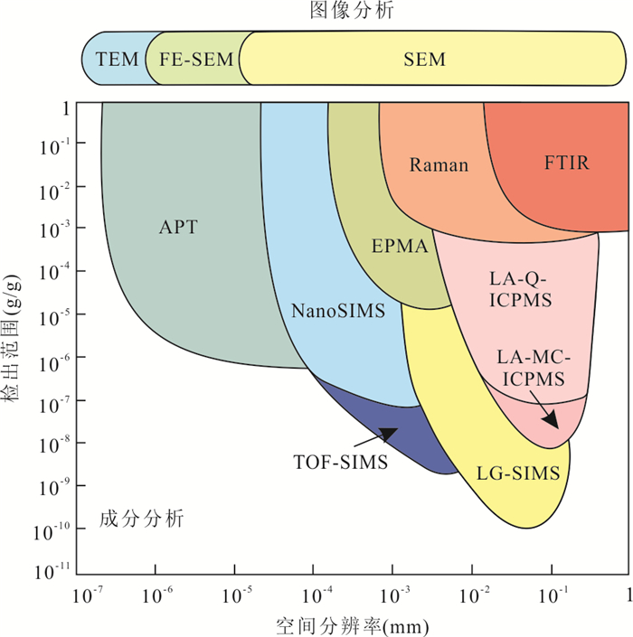
图 3 常见原位分析技术的空间分辨率和探测范围
SEM.扫描电镜;FE-SEM.场发射扫描电镜;TEM.透射电镜;FTIR.傅里叶变换红外光谱;Raman.拉曼光谱;EPMA.电子探针;LA-Q-ICPMS.激光剥蚀‒四级杆等离子体质谱;LA-MC-ICPMS.激光剥蚀‒多接收器等离子体质谱;LG-SIMS.大型离子探针;NanoSIMS.纳米离子探针;TOF-SIMS.飞行时间二次离子质谱离子探针;APT.三维原子探针.修改自Li and Li(2016)
Fig. 3. Spatial resolutions and detection ranges for common in-situ analysis techniques
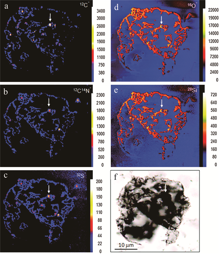
图 5 太古代Farrel石英岩中一个大球体的NanoSIMS元素分布图
a~e. NanoSIMS元素分布碳(12C‒)、氮(12C14N‒)、硫(32S‒)、氧(16O‒)以及硅(28Si‒),离子强度的变化由校准条显示,颜色越亮表示强度越高;f. 球体在透射光下的光学显微照片;白色箭头指的是具有较高的12C‒、12C14N‒、32S‒;a~f中比例尺一致;图片来源于Oehler et al.(2010)
Fig. 5. NanoSIMS element maps of a large spheroid in chert from the Farrel quartzite
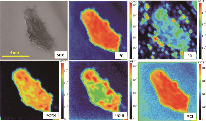
图 6 Tissint火星陨石中熔脉里面有机碳碳颗粒NanoSIMS成像
分析包括碳(C)、氢(12C1H-)、氮(12C14N-)、硫(S)、氯(Cl),离子强度的变化由校准条显示,颜色越亮表示强度越高.图片来源于Lin et al.(2014)
Fig. 6. NanoSIMS ion maps of organic carbons entrained in shock-melt veins of the Tissint meteorolite
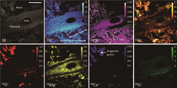
图 7 现代微生物岩中丝状蓝藻的NanoSIMS离子图像
图中显示碳(12C-)、氮(26CN-)、硫(32S-)、氧(16O-)、镁(24Mg+)、钙(40Ca+)和硅(28Si-)的分布,以及扫描电镜电子图像(SE).图像展示鞘(sheath)的纵向截面,鞘外被胞外聚合物(EPS)包围(在26CN-图像中由虚线圈出的亮区表示).离子强度的变化由校准条显示,颜色越亮表示强度越高.文石颗粒与EPS紧密关联.比例尺为20 μm.图片来源于Wacey et al.(2010)
Fig. 7. NanoSIMS ion images of the filamentous cyanobacterium
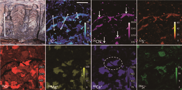
图 8 西澳大利亚约27亿年前Tumbiana组微生物岩的NanoSIMS离子图像
图中展示碳(12C-)、氮(26CN-)、硫(32S-)、氧(16O-)、镁(24Mg+)、钙(40Ca+)和硅(28Si-)的分布.左上角为微生物岩的野外照片.样品中未发现微化石,但离子图像(12C-、26CN-和32S-,以及与16O-的反相关)突显贯穿图像中心和底部的有机物(在26CN-图像中用箭头标出).离子强度的变化通过校准条表示,颜色越亮表示强度越高.此外,图像中同时存在碳酸钙和硅质颗粒,中心部分出现有机物可能捕获碳酸盐颗粒的现象(在40Ca+图像中用圆圈标出).离子图像的比例尺为10 μm;野外照片中铅笔的长度为15 cm.图片来源于Wacey et al.(2010)
Fig. 8. NanoSIMS ion images of a microbialite from the ∼2 720 Ma Tumbiana Formation, Western Australia
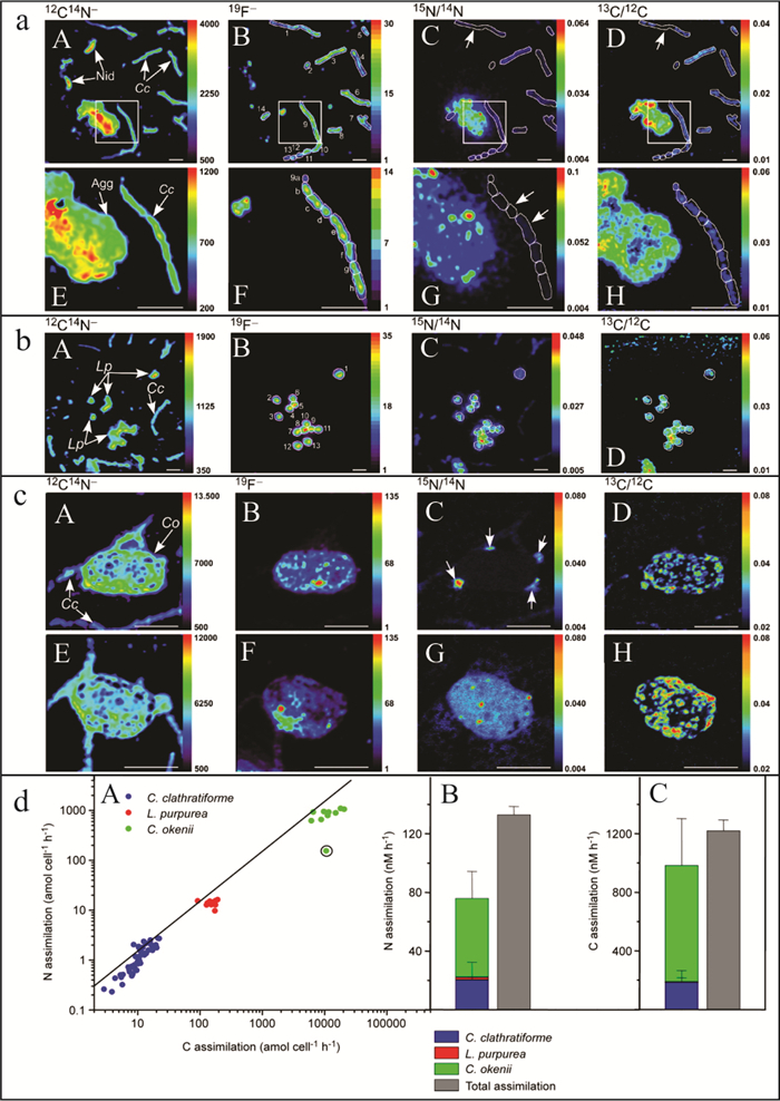
图 9 单个细胞对碳(H13CO3‒)和氮(15NH4+)的吸收平行二次离子图像(a~c),以及C. clathratiforme, L. purpurea, and C. okenii单个微生物细胞代谢活动中对碳和铵的吸收定量统计(d)
a(A~H)主要为C. clathratiforme细胞,b(A~D)主要为L. purpurea细胞,c(A~H)主要为C. okenii细胞.Cc代表C. clathratiforme,Lp代表L. purpurea,Co代表C. okenii,Nid代表未鉴定微生物,Agg代表为未鉴定的细菌的集合体,比例尺5 μm;d(A)中该线代表Redfield理论中海洋浮游植物的碳氮比约为6.6,并对总铵的贡献,d(B)为对总铵的贡献,d(C)为每个族群对系统中总溶解无机碳吸收的贡献.修改自Musat et al.(2008)
Fig. 9. Parallel secondary ion images of 15N-ammonium and 13C-inorganic carbon uptake by individual microorganism cells (a~c), and ammonium versus inorganic carbon uptake by individual C. clathratiforme, L. purpurea, and C. okenii cells (d)
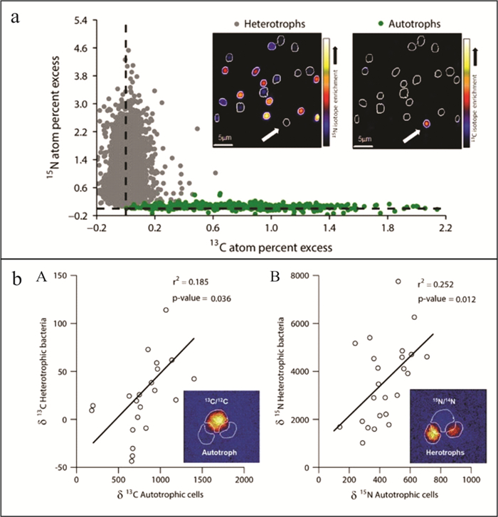
图 10 利用同位素标记的13C碳酸氢盐(bicarbonate)和15N亮氨酸(leucine)培养的微生物细胞中13C和15N同位素富集的对比(a),浮游植物细胞与附着的细菌细胞的13C掺入率之间呈正相关(b-A),细菌与附着的浮游植物细胞的15N掺入率之间呈正相关(b-B).修改自Arandia-Gorostidi et al. (2017)
图a中虚线为同位素天然丰度,每个点代表一个微生物细胞,通过整合NanoSIMS图像中定义的感兴趣区域的像素获得富集程度.NanoSIMS富集程度图像显示已鉴定的异养生物(Heterotrophs)和一个自养生物(Autotroph,白色箭头所指)
Fig. 10. Comparison between 13C and 15N isotopic enrichment for all analyzed microbial cells incubated with 13C bicarbonate and 15N leucine (a). Positive correlation between the 13C incorporation rates of phytoplankton cells and those of their attached bacterial cells, analyzed with a smaller raster for one of the replicates of the warm incubations (b-A), Similar positive correlation between the 15N incorporation rates of bacteria and those of the phytoplankton cells to which they were attached (b-B). Modified by Arandia-Gorostidi et al. (2017)
-
Allwood, A. C., Walter, M. R., Kamber, B. S., et al., 2006. Stromatolite Reef from the Early Archaean Era of Australia. Nature, 441: 714-718. https://doi.org/10.1038/nature04764 Altermann, W., Kazmierczak, J., 2003. Archean Microfossils: A Reappraisal of Early Life on Earth. Research in Microbiology, 154(9): 611-617. https://doi.org/10.1016/j.resmic.2003.08.006 Arandia-Gorostidi, N., Weber, P. K., Alonso-Sáez, L., et al., 2017. Elevated Temperature Increases Carbon and Nitrogen Fluxes between Phytoplankton and Heterotrophic Bacteria through Physical Attachment. The ISME Journal, 11(3): 641-650. https://doi.org/10.1038/ismej.2016.156 Brasier, M., McLoughlin, N., Green, O., et al., 2006. A Fresh Look at the Fossil Evidence for Early Archaean Cellular Life. Philosophical Transactions of the Royal Society B: Biological Sciences, 361(1470): 887-902. https://doi.org/10.1098/rstb.2006.1835 Bryant, R. N., Houghton, J. L., Jones, C., et al., 2023. Deconvolving Microbial and Environmental Controls on Marine Sedimentary Pyrite Sulfur Isotope Ratios. Science, 382(6673): 912-915. https://doi.org/10.1126/science.adg6103 Burne, R. V., Moore, L. S., 1987. Microbialites: Organosedimentary Deposits of Benthic Microbial Communities. Palaios, 2(3): 241. https://doi.org/10.2307/3514674 Canfield, D. E., 2001. Biogeochemistry of Sulfur Isotopes. Reviews in Mineralogy and Geochemistry, 43(1): 607-636. https://doi.org/10.2138/gsrmg.43.1.607 Chang, H. J., Chu, X. L., Feng, L. J., et al., 2009. Redox Sensitive Trace Elements as Paleoenvironments Proxies. Geological Review, 55(1): 91-99 (in Chinese with English abstract). http://search.cnki.net/down/default.aspx?filename=DZLP200901015&dbcode=CJFD&year=2009&dflag=pdfdown Chen, J., Yao, S. P., 2005. Geomicrobiology and Its Progress. Geological Journal of China Universities, 11(2): 154-166 (in Chinese with English abstract). http://en.cnki.com.cn/Article_en/CJFDTOTAL-GXDX200502001.htm Chen, Y., Hu, Z. C., Jia, L. H., et al., 2021. Progress of Microbeam Analytical Technologies in the Past Decade (2011-2020) and Prospect. Bulletin of Mineralogy, Petrology and Geochemistry, 40(1): 1-35 (in Chinese with English abstract). Chen, Z. Q., Zhou, C., Stanley, G. J., 2017. Biosedimentary Records of China from the Precambrian to Present. Palaeogeography, Palaeoclimatology, Palaeoecology, 474: 1-6. https://doi.org/10.1016/j.palaeo.2017.03.002 Chen, Z. Q., Tu, C. Y., Pei, Y., et al., 2019. Biosedimentological Features of Major Microbe-Metazoan Transitions (MMTs) from Precambrian to Cenozoic. Earth-Science Reviews, 189: 21-50. https://doi.org/10.1016/j.earscirev.2019.01.015 Green, J., Hoehler, T., Neveu, M., et al., 2021. Call for a Framework for Reporting Evidence for Life beyond Earth. Nature, 598: 575-579. https://doi.org/10.1038/s41586-021-03804-9 Grotzinger, J. P., Rothman, D. H., 1996. An Abiotic Model for Stromatolite Morphogenesis. Nature, 383: 423-425. https://doi.org/10.1038/383423a0 Guo, Z. X., Papineau, D., O'Neil, J., et al., 2024. Abiotic Synthesis of Graphitic Carbons in the Eoarchean Saglek-Hebron Metasedimentary Rocks. Nature Communications, 15: 5679. https://doi.org/10.1038/s41467-024-50134-1 Halevy, I., Fike, D. A., Pasquier, V., et al., 2023. Sedimentary Parameters Control the Sulfur Isotope Composition of Marine Pyrite. Science, 382(6673): 946-951. https://doi.org/10.1126/science.adh1215 Hansen, C. J., Castillo-Rogez, J., Grundy, W., et al., 2021. Triton: Fascinating Moon, Likely Ocean World, Compelling Destination!. The Planetary Science Journal, 2(4): 137. https://doi.org/10.3847/psj/abffd2 Hao, J. L., Zhang, L. P., Li, Z. Y., et al., 2024. Sulfur Isotopic Analysis of Sedimentary Pyrite at Micro-Nano Scale and Its Implications to Oil/Gas Generation in the Dongying Depression. Acta Petrologica Sinica, 40(1): 313-322 (in Chinese with English abstract). doi: 10.18654/1000-0569/2024.01.17 Harazim, D., Virtasalo, J. J., Denommee, K. C., et al., 2020. Exceptional Sulfur and Iron Isotope Enrichment in Millimetre-Sized, Early Palaeozoic Animal Burrows. Scientific Reports, 10: 20270. https://doi.org/10.1038/s41598-020-76296-8 Hu, H. W., Zhang, L. M., He, J. Z., 2013. Application of Nano-Scale Secondary Ion Mass Spectrometry to Microbial Ecology Study. Acta Ecologica Sinica, 33(2): 348-357 (in Chinese with English abstract). doi: 10.5846/stxb201111301826 Hu, Y. L., Wang, W., Zhao, X. Y., et al., 2025. Extreme Sulfur Isotope Heterogeneity in Individual Ediacaran Pyrite Grains Revealed by NanoSIMS Analysis. Marine and Petroleum Geology, 171: 107201. https://doi.org/10.1016/j.marpetgeo.2024.107201 Imachi, H., Nobu, M. K., Nakahara, N., et al., 2020. Isolation of an Archaeon at the Prokaryote-Eukaryote Interface. Nature, 577: 519-525. https://doi.org/10.1038/s41586-019-1916-6 Jia, X., Kivelson, M. G., 2021. The Magnetosphere of Ganymede. In: Maggiolo, R., André, N., Hasegawa, H., et al., eds., Magnetospheres in the Solar System. Wiley, Hoboken, 557-573. https://doi.org/10.1002/9781119815624.ch35 Jørgensen, B. B., Findlay, A. J., Pellerin, A., 2019. The Biogeochemical Sulfur Cycle of Marine Sediments. Frontiers in Microbiology, 10: 849. https://doi.org/10.3389/fmicb.2019.00849 Jørgensen, B. B., Kasten, S., 2006. Sulfur Cycling and Methane Oxidation. In: Schulz, H. D., Zabel, M., eds., Marine Geochemistry. Springer-Verlag, Berlin, 271-309. https://doi.org/10.1007/3-540-32144-6_8 Li, Q. L., Yang, W., Liu, Y., et al., 2013. Ion Microprobe Microanalytical Techniques and Their Applications in Earth Sciences. Bulletin of Mineralogy, Petrology and Geochemistry, 32(3): 310-327 (in Chinese with English abstract). doi: 10.3969/j.issn.1007-2802.2013.03.004 Li, W. J., Jiang, H. C., 2018. Geomicrobiology: A New Interdisciplinary Subject. Acta Microbiologica Sinica, 58(4): 521-524 (in Chinese with English abstract). Li, X. H., Li, Q. L., 2016. Major Advances in Microbeam Analytical Techniques and Their Applications in Earth Science. Science Bulletin, 61(23): 1785-1787. https://doi.org/10.1007/s11434-016-1197-5 Lin, W., Shen, J. X., Pan, Y. X., 2022. On Astrobiological Research in China. Earth Science, 47(11): 4108-4113 (in Chinese with English abstract). Lin, Y. T., El Goresy, A., Hu, S., et al., 2014. NanoSIMS Analysis of Organic Carbon from the Tissint Martian Meteorite: Evidence for the Past Existence of Subsurface Organic-Bearing Fluids on Mars. Meteoritics & Planetary Science, 49(12): 2201-2218. https://doi.org/10.1111/maps.12389 Lin, Z. Y., Sun, X. M., Strauss, H., et al., 2017. Multiple Sulfur Isotope Constraints on Sulfate-Driven Anaerobic Oxidation of Methane: Evidence from Authigenic Pyrite in Seepage Areas of the South China Sea. Geochimica et Cosmochimica Acta, 211: 153-173. https://doi.org/10.1016/j.gca.2017.05.015 Lopes, R. M. C., Kirk, R. L., Mitchell, K. L., et al., 2013. Cryovolcanism on Titan: New Results from Cassini RADAR and VIMS. Journal of Geophysical Research: Planets, 118(3): 416-435. https://doi.org/10.1002/jgre.20062 Lovelock, J. E., 1983. Gaia as Seen through the Atmosphere. In: Westbroek, P., Jong, E. W., eds., Biomineralization and Biological Metal Accumulation. Springer, Dordrecht, 15-25. https://doi.org/10.1007/978-94-009-7944-4_2 Marin-Carbonne, J., Decraene, M. N., Havas, R., et al., 2022. Early Precipitated Micropyrite in Microbialites: A Time Capsule of Microbial Sulfur Cycling. Geochemical Perspectives Letters, 21: 7-12. https://doi.org/10.7185/geochemlet.2209 Mayali, X., 2020. NanoSIMS: Microscale Quantification of Biogeochemical Activity with Large-Scale Impacts. Annual Review of Marine Science, 12: 449-467. https://doi.org/10.1146/annurev-marine-010419-010714 McLoughlin, N., Wilson, L. A., Brasier, M. D., 2008. Growth of Synthetic Stromatolites and Wrinkle Structures in the Absence of Microbes-Implications for the Early Fossil Record. Geobiology, 6(2): 95-105. https://doi.org/10.1111/j.1472-4669.2007.00141.x Moorbath, S., 2005. Dating Earliest Life. Nature, 434(7030): 155. https://doi.org/10.1038/434155a Musat, N., Halm, H., Winterholler, B., et al., 2008. A Single-Cell View on the Ecophysiology of Anaerobic Phototrophic Bacteria. Proceedings of the National Academy of Sciences, 105(46): 17861-17866. https://doi.org/10.1073/pnas.0809329105 Oehler, D. Z., Robert, F., Walter, M. R., et al., 2010. Diversity in the Archean Biosphere: New Insights from NanoSIMS. Astrobiology, 10(4): 413-424. https://doi.org/10.1089/ast.2009.0426 Papineau, D., She, Z., Dodd, M. S., et al., 2022. Metabolically Diverse Primordial Microbial Communities in Earth's Oldest Seafloor-Hydrothermal Jasper. Science Advances, 8(15): eabm2296. https://doi.org/10.1126/sciadv.abm2296 Pasquier, V., Bryant, R. N., Fike, D. A., et al., 2021. Strong Local, Not Global, Controls on Marine Pyrite Sulfur Isotopes. Science Advances, 7(9): eabb7403. https://doi.org/10.1126/sciadv.abb7403 Postberg, F., Khawaja, N., Abel, B., et al., 2018. Macromolecular Organic Compounds from the Depths of Enceladus. Nature, 558: 564-568. https://doi.org/10.1038/s41586-018-0246-4 Qiu, X. C., 2022. Geomicrobiological Process and Paleoceanographical Analysis of Early Triassic Microbialites and Fish-Bearing Calcareous Nodules in South China (Dissertation). China University of Geosciences, Wuhan (in Chinese with English abstract). Raven, M. R., Sessions, A. L., Fischer, W. W., et al., 2016. Sedimentary Pyrite δ34S Differs from Porewater Sulfide in Santa Barbara Basin: Proposed Role of Organic Sulfur. Geochimica et Cosmochimica Acta, 186: 120-134. https://doi.org/10.1016/j.gca.2016.04.037 Schopf, J. W., 2006. Fossil Evidence of Archaean Life. Philosophical Transactions of the Royal Society B: Biological Sciences, 361(1470): 869-885. https://doi.org/10.1098/rstb.2006.1834 Schuhmacher, M., Rasser, B., De Chambost, E., et al., 1999. Recent Instrumental Developments in Magnetic Sector SIMS. Fresenius' Journal of Analytical Chemistry, 365(1): 12-18. https://doi.org/10.1007/s002160051438 Steinhauser, M. L., Bailey, A. P., Senyo, S. E., et al., 2012. Multi-Isotope Imaging Mass Spectrometry Quantifies Stem Cell Division and Metabolism. Nature, 481: 516-519. https://doi.org/10.1038/nature10734 Sun, S., Wang, C. S., 2008. Gaia Theory and Earth System Science. Acta Geologica Sinica, 82(1): 1-8 (in Chinese with English abstract). doi: 10.1111/j.1755-6724.2008.tb00319.x Thomazo, C., Vennin, E., Brayard, A., et al., 2016. A Diagenetic Control on the Early Triassic Smithian-Spathian Carbon Isotopic Excursions Recorded in the Marine Settings of the Thaynes Group (Utah, USA). Geobiology, 14(3): 220-236. https://doi.org/10.1111/gbi.12174 Wacey, D., Gleeson, D., Kilburn, M. R., 2010. Microbialite Taphonomy and Biogenicity: New Insights from NanoSIMS. Geobiology, 8(5): 403-416. https://doi.org/10.1111/j.1472-4669.2010.00251.x Walter, M. R., Buick, R., Dunlop, J. S. R., 1980. Stromatolites 3, 400-3, 500 Myr Old from the North Pole Area, Western Australia. Nature, 284(5755): 443-445. https://doi.org/10.1038/284443a0 Wang, W., Hu, Y. L., Muscente, A. D., et al., 2021. Revisiting Ediacaran Sulfur Isotope Chemostratigraphy with in Situ nanoSIMS Analysis of Sedimentary Pyrite. Geology, 49(6): 611-616. https://doi.org/10.1130/G48262.1 Wu, K. J., Zhou, L., Tahon, G., et al., 2024. Isolation of a Methyl-Reducing Methanogen Outside the Euryarchaeota. Nature, 632: 1124-1130. https://doi.org/10.1038/s41586-024-07728-y Xie, S. C., Gong, Y. M., Tong, J. N., et al., 2006. A Transition from Paleontology to Geobiology. Chinese Science Bulletin, 51(19): 2327-2336 (in Chinese). doi: 10.1007/s11434-006-2111-3 Xie, S. C., Yang, H., Luo, G. M., et al., 2012. Geomicrobial Functional Groups: A Window on the Interaction between Life and Environments. Chinese Science Bulletin, 57(1): 3-22 (in Chinese). doi: 10.1360/csb2012-57-1-3 Yang, W., Hu, S., Zhang, J. C., et al., 2015. Nano SIMS Analytical Technique and Its Applications in Earth Sciences. Science in China (Series D), 45(9): 1335-1346 (in Chinese). doi: 10.1007/s11430-015-5106-6 Zhang, J. C., Lin, Y. T., Yang, W., et al., 2014. Improved Precision and Spatial Resolution of Sulfur Isotope Analysis Using NanoSIMS. Journal of Analytical Atomic Spectrometry, 29(10): 1934-1943. https://doi.org/10.1039/C4JA00140K 常华进, 储雪蕾, 冯连君, 等, 2009. 氧化还原敏感微量元素对古海洋沉积环境的指示意义. 地质论评, 55(1): 91-99. 陈骏, 姚素平, 2005. 地质微生物学及其发展方向. 高校地质学报, 11(2): 154-166. 陈意, 胡兆初, 贾丽辉, 等, 2021. 微束分析测试技术十年(2011—2020)进展与展望. 矿物岩石地球化学通报, 40(1): 1-35. 郝佳龙, 张刘平, 李照阳, 等, 2024. 矿物微‒纳尺度硫同位素分析及在东营凹陷油气成藏的应用示范. 岩石学报, 40(1): 313-322. 胡行伟, 张丽梅, 贺纪正, 2013. 纳米二次离子质谱技术(NanoSIMS)在微生物生态学研究中的应用. 生态学报, 33(2): 348-357. 李秋立, 杨蔚, 刘宇, 等, 2013. 离子探针微区分析技术及其在地球科学中的应用进展. 矿物岩石地球化学通报, 32(3): 310-327. 李文均, 蒋宏忱, 2018. 地质微生物学: 一门新兴的交叉学科. 微生物学报, 58(4): 521-524. 林巍, 申建勋, 潘永信, 2022. 关于我国天体生物学研究的思考. 地球科学, 47(11): 4108-4113. 仇鑫程, 2022. 华南早三叠世微生物岩和钙质鱼结核的微生物地质过程和古海洋环境分析(博士学位论文). 武汉: 中国地质大学. 孙枢, 王成善, 2008. Gaia理论与地球系统科学. 地质学报, 82(1): 1-8. 谢树成, 龚一鸣, 童金南, 等, 2006. 从古生物学到地球生物学的跨越. 科学通报, 51(19): 2327-2336. 谢树成, 杨欢, 罗根明, 等, 2012. 地质微生物功能群: 生命与环境相互作用的重要突破口. 科学通报, 57(1): 3-22. 杨蔚, 胡森, 张建超, 等, 2015. 纳米离子探针分析技术及其在地球科学中的应用. 中国科学(D辑), 45(9): 1335-1346. -

 下载:
下载:
 点击查看大图
计量
- 文章访问数: 211
- HTML全文浏览量: 83
- PDF下载量: 42
- 被引次数: 0
